Crystal Engineering of Self-Assembled Porous Protein Materials in Living Cells
Abe, S., Tabe, H., Ijiri, H., Yamashita, K., Hirata, K., Atsumi, K., Shimoi, T., Akai, M., Mori, H., Kitagawa, S., Ueno, T.(2017) ACS Nano 11: 2410-2419
- PubMed: 28094987
- DOI: https://doi.org/10.1021/acsnano.6b06099
- Primary Citation of Related Structures:
5GQI, 5GQJ, 5GQK, 5GQL, 5GQM, 5GQN - PubMed Abstract:
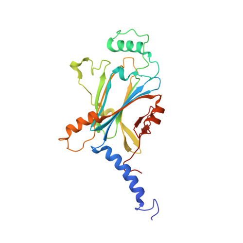
Crystalline porous materials have been investigated for development of important applications in molecular storage, separations, and catalysis. The potential of protein crystals is increasing as they become better understood. Protein crystals have been regarded as porous materials because they present highly ordered 3D arrangements of protein molecules with high porosity and wide range of pore sizes. However, it remains difficult to functionalize protein crystals in living cells. Here, we report that polyhedra, a natural crystalline protein assembly of polyhedrin monomer (PhM) produced in insect cells infected by cypovirus, can be engineered to extend porous networks by deleting selected amino acid residues located on the intermolecular contact region of PhM. The adsorption rates and quantities of fluorescent dyes stored within the mutant crystals are increased relative to those of the wild-type polyhedra crystal (WTPhC) under both in vitro and in vivo conditions. These results provide a strategy for designing self-assembled protein materials with applications in molecular recognition and storage of exogenous substances in living cell as well as an entry point for development of bioorthogonal chemistry and in vivo crystal structure analysis.
Organizational Affiliation:
School of Life Science and Technology, Tokyo Institute of Technology , Nagatsuta-cho, Midori-ku, Yokohama 226-8501, Japan.