DNA polymorphism in crystals: three stable conformations for the decadeoxynucleotide d(GCATGCATGC).
Thirugnanasambandam, A., Karthik, S., Artheswari, G., Gautham, N.(2016) Acta Crystallogr D Struct Biol 72: 780-788
- PubMed: 27303798
- DOI: https://doi.org/10.1107/S2059798316006306
- Primary Citation of Related Structures:
5FHJ, 5FHL - PubMed Abstract:
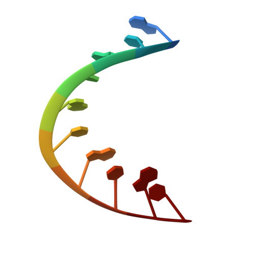
High-resolution structures of DNA fragments determined using X-ray crystallography or NMR have provided descriptions of a veritable alphabet of conformations. They have also shown that DNA is a flexible molecule, with some sequences capable of adopting two different structures. Here, the first example is presented of a DNA fragment that can assume three different and distinct conformations in crystals. The decanucleotide d(GCATGCATGC) was previously reported to assume a single-stranded double-fold structure. In one of the two crystal structures described here the decamer assumes both the double-fold conformation and, simultaneously, the more conventional B-type double-helical structure. In the other crystal the sequence assumes the A-type double-helical conformation. These results, taken together with CD spectra, which were recorded as the decamer was titrated against four metal ions and spermine, indicate that the molecule may exist as a mixed population of structures in solution. Small differences in the environmental conditions, such as the concentration of metal ion, may decide which of these crystallizes out. The results also support the idea that it may be possible for DNA to change its structure to suit the binding requirements of proteins or drugs.
Organizational Affiliation:
CAS in Crystallography and Biophysics, University of Madras, Guindy Campus, Chennai 600 025, India.