5FCN

microtubule binding domain of human CEP135


Experimental Data Snapshot

  • Method: X-RAY DIFFRACTION
  • Resolution: 1.80 Å
  • R-Value Free: 0.238 
  • R-Value Work: 0.216 
  • R-Value Observed: 0.217 

wwPDB Validation   3D Report Full Report


This is version 1.3 of the entry. See complete history


Literature

The Human Centriolar Protein CEP135 Contains a Two-Stranded Coiled-Coil Domain Critical for Microtubule Binding.

Kraatz, S.Guichard, P.Obbineni, J.M.Olieric, N.Hatzopoulos, G.N.Hilbert, M.Sen, I.Missimer, J.Gonczy, P.Steinmetz, M.O.

(2016) Structure 24: 1358-1371

  • DOI: https://doi.org/10.1016/j.str.2016.06.011
  • Primary Citation of Related Structures:  
    5FCM, 5FCN

  • PubMed Abstract: 

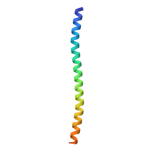
    Centrioles are microtubule-based structures that play important roles notably in cell division and cilium biogenesis. CEP135/Bld10p family members are evolutionarily conserved microtubule-binding proteins important for centriole formation. Here, we analyzed in detail the microtubule-binding activity of human CEP135 (HsCEP135). X-ray crystallography and small-angle X-ray scattering in combination with molecular modeling revealed that the 158 N-terminal residues of HsCEP135 (HsCEP135-N) form a parallel two-stranded coiled-coil structure. Biochemical, cryo-electron, and fluorescence microscopy analyses revealed that in vitro HsCEP135-N interacts with tubulin, protofilaments, and microtubules and induces the formation of microtubule bundles. We further identified a 13 amino acid segment spanning residues 96-108, which represents a major microtubule-binding site in HsCEP135-N. Within this segment, we identified a cluster of three lysine residues that contribute to the microtubule bundling activity of HsCEP135-N. Our results provide the first structural information on CEP135/Bld10p proteins and offer insights into their microtubule-binding mechanism.


  • Organizational Affiliation

    Laboratory of Biomolecular Research, Department of Biology and Chemistry, Paul Scherrer Institut, 5232 Villigen, Switzerland.


Macromolecules
Find similar proteins by:  (by identity cutoff)  |  3D Structure
Entity ID: 1
MoleculeChains Sequence LengthOrganismDetailsImage
Centrosomal protein of 135 kDaA [auth B],
B [auth A]
65Homo sapiensMutation(s): 0 
Gene Names: CEP135CEP4KIAA0635
UniProt & NIH Common Fund Data Resources
Find proteins for Q66GS9 (Homo sapiens)
Explore Q66GS9 
Go to UniProtKB:  Q66GS9
PHAROS:  Q66GS9
GTEx:  ENSG00000174799 
Entity Groups  
Sequence Clusters30% Identity50% Identity70% Identity90% Identity95% Identity100% Identity
UniProt GroupQ66GS9
Sequence Annotations
Expand
  • Reference Sequence
Experimental Data & Validation

Experimental Data

  • Method: X-RAY DIFFRACTION
  • Resolution: 1.80 Å
  • R-Value Free: 0.238 
  • R-Value Work: 0.216 
  • R-Value Observed: 0.217 
  • Space Group: I 41 2 2
Unit Cell:
Length ( Å )Angle ( ˚ )
a = 78.85α = 90
b = 78.85β = 90
c = 95.92γ = 90
Software Package:
Software NamePurpose
PHENIXrefinement
XDSdata reduction
XSCALEdata scaling
PHASERphasing

Structure Validation

View Full Validation Report


Entry History & Funding Information

Deposition Data


Funding OrganizationLocationGrant Number
Swiss National Science FoundationSwitzerland310030B_138659

Revision History  (Full details and data files)

  • Version 1.0: 2016-07-20
    Type: Initial release
  • Version 1.1: 2016-08-10
    Changes: Database references
  • Version 1.2: 2017-10-18
    Changes: Database references
  • Version 1.3: 2024-01-10
    Changes: Data collection, Database references, Refinement description