5FBF

S1 nuclease from Aspergillus oryzae in complex with two molecules of 2'-deoxycytidine-5'-monophosphate

  • Classification: HYDROLASE
  • Organism(s): Aspergillus oryzae RIB40
  • Expression System: Aspergillus oryzae
  • Mutation(s): No 

  • Deposited: 2015-12-14 Released: 2016-12-28 
  • Deposition Author(s): Koval, T., Oestergaard, L.H., Dohnalek, J.
  • Funding Organization(s): Ministry of Education, Youth and Sports of the Czech Republic, BIOCEV: Biotechnology and Biomedicine Centre of the Academy of Sciences and Charles University from the European Regional Development Fund, European Community

Experimental Data Snapshot

  • Method: X-RAY DIFFRACTION
  • Resolution: 1.04 Å
  • R-Value Free: 0.135 
  • R-Value Work: 0.111 
  • R-Value Observed: 0.113 

wwPDB Validation   3D Report Full Report


Ligand Structure Quality Assessment 


This is version 1.2 of the entry. See complete history


Literature

Structural and Catalytic Properties of S1 Nuclease from Aspergillus oryzae Responsible for Substrate Recognition, Cleavage, Non-Specificity, and Inhibition.

Koval, T.stergaard, L.H.Lehmbeck, J.Nrgaard, A.Lipovova, P.Duskova, J.Skalova, T.Trundova, M.Kolenko, P.Fejfarova, K.Stransky, J.Svecova, L.Hasek, J.Dohnalek, J.

(2016) PLoS One 11: e0168832-e0168832

  • DOI: https://doi.org/10.1371/journal.pone.0168832
  • Primary Citation of Related Structures:  
    5FB9, 5FBA, 5FBB, 5FBC, 5FBD, 5FBF, 5FBG

  • PubMed Abstract: 

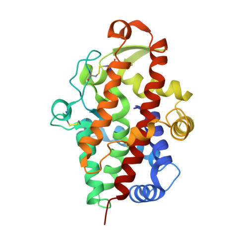
    The single-strand-specific S1 nuclease from Aspergillus oryzae is an archetypal enzyme of the S1-P1 family of nucleases with a widespread use for biochemical analyses of nucleic acids. We present the first X-ray structure of this nuclease along with a thorough analysis of the reaction and inhibition mechanisms and of its properties responsible for identification and binding of ligands. Seven structures of S1 nuclease, six of which are complexes with products and inhibitors, and characterization of catalytic properties of a wild type and mutants reveal unknown attributes of the S1-P1 family. The active site can bind phosphate, nucleosides, and nucleotides in several distinguished ways. The nucleoside binding site accepts bases in two binding modes-shallow and deep. It can also undergo remodeling and so adapt to different ligands. The amino acid residue Asp65 is critical for activity while Asn154 secures interaction with the sugar moiety, and Lys68 is involved in interactions with the phosphate and sugar moieties of ligands. An additional nucleobase binding site was identified on the surface, which explains the absence of the Tyr site known from P1 nuclease. For the first time ternary complexes with ligands enable modeling of ssDNA binding in the active site cleft. Interpretation of the results in the context of the whole S1-P1 nuclease family significantly broadens our knowledge regarding ligand interaction modes and the strategies of adjustment of the enzyme surface and binding sites to achieve particular specificity.


  • Organizational Affiliation

    Laboratory of Structure and Function of Biomolecules, Institute of Biotechnology CAS, v. v. i., Biocev, Vestec, Czech Republic.


Macromolecules
Find similar proteins by:  (by identity cutoff)  |  3D Structure
Entity ID: 1
MoleculeChains Sequence LengthOrganismDetailsImage
Nuclease S1267Aspergillus oryzae RIB40Mutation(s): 0 
Gene Names: nucSAO090001000075
EC: 3.1.30.1
UniProt
Find proteins for P24021 (Aspergillus oryzae (strain ATCC 42149 / RIB 40))
Explore P24021 
Go to UniProtKB:  P24021
Entity Groups  
Sequence Clusters30% Identity50% Identity70% Identity90% Identity95% Identity100% Identity
UniProt GroupP24021
Sequence Annotations
Expand
  • Reference Sequence
Small Molecules
Ligands 4 Unique
IDChains Name / Formula / InChI Key2D Diagram3D Interactions
DCM
Query on DCM

Download Ideal Coordinates CCD File 
G [auth A],
H [auth A]
2'-DEOXYCYTIDINE-5'-MONOPHOSPHATE
C9 H14 N3 O7 P
NCMVOABPESMRCP-SHYZEUOFSA-N
NAG
Query on NAG

Download Ideal Coordinates CCD File 
E [auth A],
F [auth A]
2-acetamido-2-deoxy-beta-D-glucopyranose
C8 H15 N O6
OVRNDRQMDRJTHS-FMDGEEDCSA-N
ZN
Query on ZN

Download Ideal Coordinates CCD File 
B [auth A],
C [auth A],
D [auth A]
ZINC ION
Zn
PTFCDOFLOPIGGS-UHFFFAOYSA-N
NA
Query on NA

Download Ideal Coordinates CCD File 
I [auth A]SODIUM ION
Na
FKNQFGJONOIPTF-UHFFFAOYSA-N
Experimental Data & Validation

Experimental Data

  • Method: X-RAY DIFFRACTION
  • Resolution: 1.04 Å
  • R-Value Free: 0.135 
  • R-Value Work: 0.111 
  • R-Value Observed: 0.113 
  • Space Group: P 21 21 21
Unit Cell:
Length ( Å )Angle ( ˚ )
a = 53.742α = 90
b = 62.388β = 90
c = 62.762γ = 90
Software Package:
Software NamePurpose
REFMACrefinement
XDSdata reduction
XDSdata scaling
MOLREPphasing
Cootmodel building

Structure Validation

View Full Validation Report


Ligand Structure Quality Assessment 


Entry History & Funding Information

Deposition Data


Funding OrganizationLocationGrant Number
Ministry of Education, Youth and Sports of the Czech RepublicCzech RepublicLG14009
BIOCEV: Biotechnology and Biomedicine Centre of the Academy of Sciences and Charles University from the European Regional Development FundCzech RepublicCZ.1.05/1.1.00/02.0109
European Community283570/8787

Revision History  (Full details and data files)

  • Version 1.0: 2016-12-28
    Type: Initial release
  • Version 1.1: 2017-01-11
    Changes: Database references
  • Version 1.2: 2020-07-29
    Type: Remediation
    Reason: Carbohydrate remediation
    Changes: Author supporting evidence, Data collection, Derived calculations, Structure summary