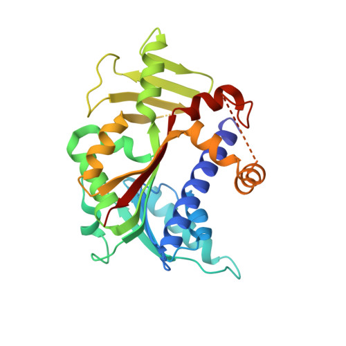
5E9W

Crystal structure of mRNA cap guanine-N7 methyltransferase obtained by limited proteolysis


Experimental Data Snapshot

  • Method: X-RAY DIFFRACTION
  • Resolution: 2.28 Å
  • R-Value Free: 0.255 
  • R-Value Work: 0.220 
  • R-Value Observed: 0.222 

wwPDB Validation   3D Report Full Report


Ligand Structure Quality Assessment 


This is version 1.6 of the entry. See complete history


Literature

Molecular basis of RNA guanine-7 methyltransferase (RNMT) activation by RAM.

Varshney, D.Petit, A.P.Bueren-Calabuig, J.A.Jansen, C.Fletcher, D.A.Peggie, M.Weidlich, S.Scullion, P.Pisliakov, A.V.Cowling, V.H.

(2016) Nucleic Acids Res 44: 10423-10436

  • DOI: https://doi.org/10.1093/nar/gkw637
  • Primary Citation of Related Structures:  
    5E8J, 5E9J, 5E9W

  • PubMed Abstract: 

    Maturation and translation of mRNA in eukaryotes requires the addition of the 7-methylguanosine cap. In vertebrates, the cap methyltransferase, RNA guanine-7 methyltransferase (RNMT), has an activating subunit, RNMT-Activating Miniprotein (RAM). Here we report the first crystal structure of the human RNMT in complex with the activation domain of RAM. A relatively unstructured and negatively charged RAM binds to a positively charged surface groove on RNMT, distal to the active site. This results in stabilisation of a RNMT lobe structure which co-evolved with RAM and is required for RAM binding. Structure-guided mutagenesis and molecular dynamics simulations reveal that RAM stabilises the structure and positioning of the RNMT lobe and the adjacent α-helix hinge, resulting in optimal positioning of helix A which contacts substrates in the active site. Using biophysical and biochemical approaches, we observe that RAM increases the recruitment of the methyl donor, AdoMet (S-adenosyl methionine), to RNMT. Thus we report the mechanism by which RAM allosterically activates RNMT, allowing it to function as a molecular rheostat for mRNA cap methylation.


  • Organizational Affiliation

    Centre for Gene Regulation and Expression, School of Life Sciences, University of Dundee, Dow Street, Dundee DD1 5EH, UK.


Macromolecules
Find similar proteins by:  (by identity cutoff)  |  3D Structure
Entity ID: 1
MoleculeChains Sequence LengthOrganismDetailsImage
mRNA cap guanine-N7 methyltransferase
A, B, C, D
312Homo sapiensMutation(s): 0 
Gene Names: RNMTKIAA0398
EC: 2.1.1.56
UniProt & NIH Common Fund Data Resources
Find proteins for O43148 (Homo sapiens)
Explore O43148 
Go to UniProtKB:  O43148
PHAROS:  O43148
GTEx:  ENSG00000101654 
Entity Groups  
Sequence Clusters30% Identity50% Identity70% Identity90% Identity95% Identity100% Identity
UniProt GroupO43148
Sequence Annotations
Expand
  • Reference Sequence
Experimental Data & Validation

Experimental Data

  • Method: X-RAY DIFFRACTION
  • Resolution: 2.28 Å
  • R-Value Free: 0.255 
  • R-Value Work: 0.220 
  • R-Value Observed: 0.222 
  • Space Group: P 21 21 21
Unit Cell:
Length ( Å )Angle ( ˚ )
a = 77.458α = 90
b = 99.157β = 90
c = 167.614γ = 90
Software Package:
Software NamePurpose
PHENIXrefinement
XDSdata reduction
SCALAdata scaling
MOLREPphasing

Structure Validation

View Full Validation Report


Ligand Structure Quality Assessment 


Entry History 

Deposition Data

Revision History  (Full details and data files)

  • Version 1.0: 2016-07-13
    Type: Initial release
  • Version 1.1: 2016-08-03
    Changes: Database references
  • Version 1.2: 2016-12-14
    Changes: Database references
  • Version 1.3: 2019-02-20
    Changes: Advisory, Data collection, Derived calculations
  • Version 1.4: 2019-06-12
    Changes: Data collection, Structure summary
  • Version 1.5: 2019-08-21
    Changes: Data collection
  • Version 1.6: 2024-01-10
    Changes: Data collection, Database references, Refinement description