


Crystal structure of Putative beta-glucanase (Rv0315 ortholog) from Mycobacterium abcesus
Abendroth, J., Davies, D.R., Lorimer, D.D., Edwards, T.E.To be published.
Experimental Data Snapshot
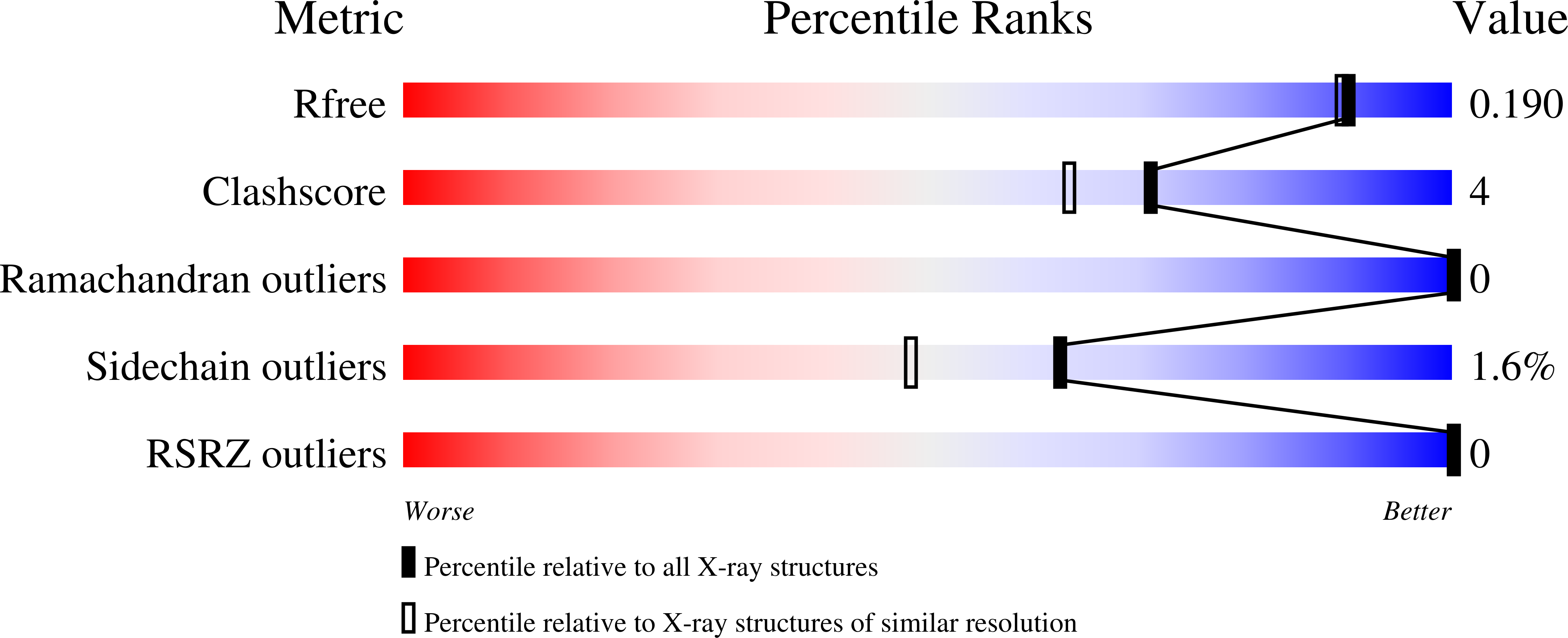
wwPDB Validation 3D Report Full Report
Entity ID: 1 | |||||
|---|---|---|---|---|---|
| Molecule | Chains | Sequence Length | Organism | Details | Image |
| Putative beta-glucanase | 257 | Mycobacteroides abscessus ATCC 19977 | Mutation(s): 0 Gene Names: MAB_1840c | 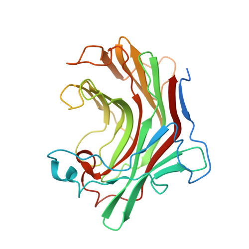 | |
UniProt | |||||
Find proteins for B1MNM2 (Mycobacteroides abscessus (strain ATCC 19977 / DSM 44196 / CCUG 20993 / CIP 104536 / JCM 13569 / NCTC 13031 / TMC 1543 / L948)) Explore B1MNM2 Go to UniProtKB: B1MNM2 | |||||
Entity Groups | |||||
| Sequence Clusters | 30% Identity50% Identity70% Identity90% Identity95% Identity100% Identity | ||||
| UniProt Group | B1MNM2 | ||||
Sequence AnnotationsExpand | |||||
| |||||
| Ligands 1 Unique | |||||
|---|---|---|---|---|---|
| ID | Chains | Name / Formula / InChI Key | 2D Diagram | 3D Interactions | |
| NA Query on NA | C [auth A], D [auth B] | SODIUM ION Na FKNQFGJONOIPTF-UHFFFAOYSA-N |  | ||
| Length ( Å ) | Angle ( ˚ ) |
|---|---|
| a = 52.39 | α = 90 |
| b = 52.39 | β = 90 |
| c = 223.35 | γ = 90 |
| Software Name | Purpose |
|---|---|
| XDS | data reduction |
| XSCALE | data scaling |
| PHASER | phasing |
| ARP | model building |
| PHENIX | refinement |
| PDB_EXTRACT | data extraction |
RCSB PDB (citation) is hosted by
RCSB PDB is a member of the

