5DJE

Crystal structure of the zuotin homology domain (ZHD) from yeast Zuo1


Experimental Data Snapshot

  • Method: X-RAY DIFFRACTION
  • Resolution: 1.85 Å
  • R-Value Free: 0.220 
  • R-Value Work: 0.174 
  • R-Value Observed: 0.176 

wwPDB Validation   3D Report Full Report


This is version 1.4 of the entry. See complete history


Literature

Dual interaction of the Hsp70 J-protein cochaperone Zuotin with the 40S and 60S ribosomal subunits.

Lee, K.Sharma, R.Shrestha, O.K.Bingman, C.A.Craig, E.A.

(2016) Nat Struct Mol Biol 23: 1003-1010

  • DOI: https://doi.org/10.1038/nsmb.3299
  • Primary Citation of Related Structures:  
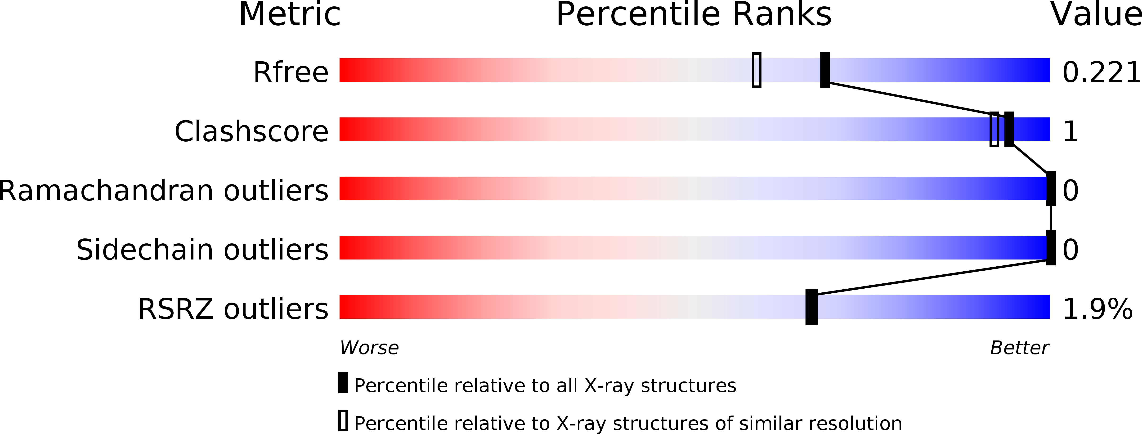
    5DJE

  • PubMed Abstract: 

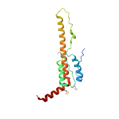
    Ribosome-associated J protein-Hsp70 chaperones promote nascent-polypeptide folding and normal translational fidelity. The J protein Zuo1 is known to span the ribosomal subunits, but understanding of its function is limited. Here we present new structural and cross-linking data allowing more precise positioning of Saccharomyces cerevisiae Zuo1 near the 60S polypeptide-exit site and suggesting interactions of Zuo1 with the ribosomal protein eL31 and 25S rRNA helix 24. The junction between the 60S-interacting and subunit-spanning helices is a hinge that positions Zuo1 on the 40S yet accommodates subunit rotation. Interaction between the Zuo1 C terminus and 40S occurs via 18S rRNA expansion segment 12 (ES12) of helix 44, which originates at the decoding site. Deletions in either ES12 or the Zuo1 C terminus alter readthrough of stop codons and -1 frameshifting. Our study offers insight into how this cotranslational chaperone system may monitor decoding-site activity and nascent-polypeptide transit, thereby coordinating protein translation and folding.


  • Organizational Affiliation

    Department of Biochemistry, University of Wisconsin-Madison, Madison, Wisconsin, USA.


Macromolecules
Find similar proteins by:  (by identity cutoff)  |  3D Structure
Entity ID: 1
MoleculeChains Sequence LengthOrganismDetailsImage
Zuotin
A, B
140Saccharomyces cerevisiae S288CMutation(s): 0 
Gene Names: ZUO1YGR285C
UniProt
Find proteins for P32527 (Saccharomyces cerevisiae (strain ATCC 204508 / S288c))
Explore P32527 
Go to UniProtKB:  P32527
Entity Groups  
Sequence Clusters30% Identity50% Identity70% Identity90% Identity95% Identity100% Identity
UniProt GroupP32527
Sequence Annotations
Expand
  • Reference Sequence
Small Molecules
Modified Residues  1 Unique
IDChains TypeFormula2D DiagramParent
MSE
Query on MSE
A, B
L-PEPTIDE LINKINGC5 H11 N O2 SeMET
Experimental Data & Validation

Experimental Data

  • Method: X-RAY DIFFRACTION
  • Resolution: 1.85 Å
  • R-Value Free: 0.220 
  • R-Value Work: 0.174 
  • R-Value Observed: 0.176 
  • Space Group: P 21 21 21
Unit Cell:
Length ( Å )Angle ( ˚ )
a = 60.48α = 90
b = 70.26β = 90
c = 95.35γ = 90
Software Package:
Software NamePurpose
PHENIXrefinement
XDSdata reduction
XSCALEdata scaling
PHASERphasing
Cootmodel building

Structure Validation

View Full Validation Report


Entry History & Funding Information

Deposition Data


Funding OrganizationLocationGrant Number
National Institutes of Health/National Institute of General Medical Sciences (NIH/NIGMS)United StatesGM31107
National Institutes of Health/National Institute of General Medical Sciences (NIH/NIGMS)United StatesU01GM098248
National Institutes of Health/National Institute of General Medical Sciences (NIH/NIGMS)United StatesU54GM094584
National Institutes of Health/National Institute of General Medical Sciences (NIH/NIGMS)United StatesU01GM094622

Revision History  (Full details and data files)

  • Version 1.0: 2016-09-28
    Type: Initial release
  • Version 1.1: 2016-10-19
    Changes: Database references
  • Version 1.2: 2016-11-16
    Changes: Database references
  • Version 1.3: 2017-09-27
    Changes: Author supporting evidence, Derived calculations
  • Version 1.4: 2019-12-25
    Changes: Author supporting evidence