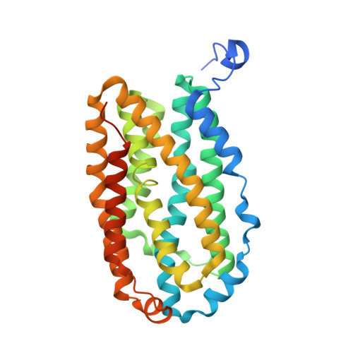
Structural Basis for Oxygen Activation at a Heterodinuclear Manganese/Iron Cofactor.
Griese, J.J., Kositzki, R., Schrapers, P., Branca, R.M., Nordstrom, A., Lehtio, J., Haumann, M., Hogbom, M.(2015) J Biol Chem 290: 25254-25272
- PubMed: 26324712
- DOI: https://doi.org/10.1074/jbc.M115.675223
- Primary Citation of Related Structures:
4XB9, 4XBV, 4XBW, 5DCO, 5DCR, 5DCS - PubMed Abstract:
Two recently discovered groups of prokaryotic di-metal carboxylate proteins harbor a heterodinuclear Mn/Fe cofactor. These are the class Ic ribonucleotide reductase R2 proteins and a group of oxidases that are found predominantly in pathogens and extremophiles, called R2-like ligand-binding oxidases (R2lox). We have recently shown that the Mn/Fe cofactor of R2lox self-assembles from Mn(II) and Fe(II) in vitro and catalyzes formation of a tyrosine-valine ether cross-link in the protein scaffold (Griese, J. J., Roos, K., Cox, N., Shafaat, H. S., Branca, R. M., Lehtiö, J., Gräslund, A., Lubitz, W., Siegbahn, P. E., and Högbom, M. (2013) Proc. Natl. Acad. Sci. U.S.A. 110, 17189-17194). Here, we present a detailed structural analysis of R2lox in the nonactivated, reduced, and oxidized resting Mn/Fe- and Fe/Fe-bound states, as well as the nonactivated Mn/Mn-bound state. X-ray crystallography and x-ray absorption spectroscopy demonstrate that the active site ligand configuration of R2lox is essentially the same regardless of cofactor composition. Both the Mn/Fe and the diiron cofactor activate oxygen and catalyze formation of the ether cross-link, whereas the dimanganese cluster does not. The structures delineate likely routes for gated oxygen and substrate access to the active site that are controlled by the redox state of the cofactor. These results suggest that oxygen activation proceeds via similar mechanisms at the Mn/Fe and Fe/Fe center and that R2lox proteins might utilize either cofactor in vivo based on metal availability.
Organizational Affiliation:
From the Stockholm Center for Biomembrane Research, Department of Biochemistry and Biophysics, Stockholm University, SE-106 91 Stockholm, Sweden.


