5D7B

Trigonal Crystal Structure of an acetylester hydrolase from Corynebacterium glutamicum


Experimental Data Snapshot

  • Method: X-RAY DIFFRACTION
  • Resolution: 3.20 Å
  • R-Value Free: 0.201 
  • R-Value Work: 0.187 
  • R-Value Observed: 0.187 

wwPDB Validation   3D Report Full Report


This is version 1.3 of the entry. See complete history


Literature

A novel esterase subfamily with alpha / beta-hydrolase fold suggested by structures of two bacterial enzymes homologous to l-homoserine O-acetyl transferases.

Tolzer, C.Pal, S.Watzlawick, H.Altenbuchner, J.Niefind, K.

(2016) FEBS Lett 590: 174-184

  • DOI: https://doi.org/10.1002/1873-3468.12031
  • Primary Citation of Related Structures:  
    5D6O, 5D7B, 5E4Y, 5EFZ

  • PubMed Abstract: 

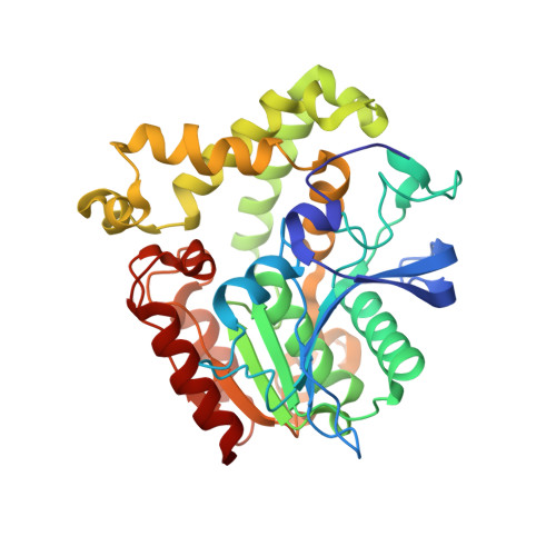
    MekB from Pseudomonas veronii and CgHle from Corynebacteriumglutamicum belong to the superfamily of α/β-hydrolase fold proteins. Based on sequence comparisons, they are annotated as homoserine transacetylases in popular databases like UNIPROT, PFAM or ESTHER. However, experimentally, MekB and CgHle were shown to be esterases that hydrolyse preferentially acetic acid esters. We describe the x-ray structures of these enzymes solved to high resolution. The overall structures confirm the close relatedness to experimentally validated homoserine acetyl transferases, but simultaneously the structures exclude the ability of MekB and CgHle to bind homoserine and acetyl-CoA. Insofar the MekB and CgHle structures suggest dividing the homoserine transacetylase family into subfamilies, namely genuine acetyl transferases and acetyl esterases with MekB and CgHle as constituting members of the latter.


  • Organizational Affiliation

    Department für Chemie, Institut für Biochemie, Universität zu Köln, Germany.


Macromolecules
Find similar proteins by:  (by identity cutoff)  |  3D Structure
Entity ID: 1
MoleculeChains Sequence LengthOrganismDetailsImage
Homoserine O-acetyltransferase
A, B
349Corynebacterium glutamicumMutation(s): 0 
Gene Names: cg0961Cgl0839
EC: 2.3.1 (PDB Primary Data), 2.3.1.31 (PDB Primary Data)
UniProt
Find proteins for Q8NS43 (Corynebacterium glutamicum (strain ATCC 13032 / DSM 20300 / BCRC 11384 / JCM 1318 / LMG 3730 / NCIMB 10025))
Explore Q8NS43 
Go to UniProtKB:  Q8NS43
Entity Groups  
Sequence Clusters30% Identity50% Identity70% Identity90% Identity95% Identity100% Identity
UniProt GroupQ8NS43
Sequence Annotations
Expand
  • Reference Sequence
Small Molecules
Ligands 1 Unique
IDChains Name / Formula / InChI Key2D Diagram3D Interactions
GOL
Query on GOL

Download Ideal Coordinates CCD File 
C [auth A]
D [auth A]
E [auth A]
F [auth A]
G [auth A]
C [auth A],
D [auth A],
E [auth A],
F [auth A],
G [auth A],
H [auth A],
I [auth B],
J [auth B],
K [auth B],
L [auth B],
M [auth B],
N [auth B],
O [auth B],
P [auth B],
Q [auth B]
GLYCEROL
C3 H8 O3
PEDCQBHIVMGVHV-UHFFFAOYSA-N
Experimental Data & Validation

Experimental Data

  • Method: X-RAY DIFFRACTION
  • Resolution: 3.20 Å
  • R-Value Free: 0.201 
  • R-Value Work: 0.187 
  • R-Value Observed: 0.187 
  • Space Group: P 31 2 1
Unit Cell:
Length ( Å )Angle ( ˚ )
a = 143.393α = 90
b = 143.393β = 90
c = 197.783γ = 120
Software Package:
Software NamePurpose
PHENIXrefinement
XDSdata reduction
Aimlessdata scaling
PHENIXphasing

Structure Validation

View Full Validation Report


Entry History & Funding Information

Deposition Data


Funding OrganizationLocationGrant Number
German Research FoundationGermanySFB 635

Revision History  (Full details and data files)

  • Version 1.0: 2015-12-16
    Type: Initial release
  • Version 1.1: 2016-02-03
    Changes: Database references
  • Version 1.2: 2017-09-06
    Changes: Author supporting evidence
  • Version 1.3: 2024-01-10
    Changes: Data collection, Database references, Refinement description