


Crystal structure of the periplasmic region of MacB from E. coli
Ha, N.C., Kim, J.S.To be published.
Experimental Data Snapshot
wwPDB Validation 3D Report Full Report

Entity ID: 1 | |||||
|---|---|---|---|---|---|
| Molecule | Chains | Sequence Length | Organism | Details | Image |
| Macrolide export ATP-binding/permease protein MacB | 226 | Escherichia coli 536 | Mutation(s): 0 Gene Names: macB, ECP_0894 EC: 3.6.3 | 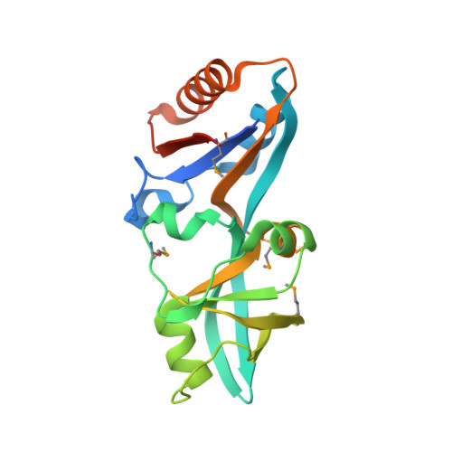 | |
UniProt | |||||
Find proteins for Q0TJH0 (Escherichia coli O6:K15:H31 (strain 536 / UPEC)) Explore Q0TJH0 Go to UniProtKB: Q0TJH0 | |||||
Entity Groups | |||||
| Sequence Clusters | 30% Identity50% Identity70% Identity90% Identity95% Identity100% Identity | ||||
| UniProt Group | Q0TJH0 | ||||
Sequence AnnotationsExpand | |||||
| |||||
| Modified Residues 1 Unique | |||||
|---|---|---|---|---|---|
| ID | Chains | Type | Formula | 2D Diagram | Parent |
| MSE Query on MSE | A, B, C, D, E A, B, C, D, E, F, G | L-PEPTIDE LINKING | C5 H11 N O2 Se |  | MET |
| Length ( Å ) | Angle ( ˚ ) |
|---|---|
| a = 67.144 | α = 90 |
| b = 78.565 | β = 99.72 |
| c = 137.922 | γ = 90 |
| Software Name | Purpose |
|---|---|
| PHENIX | refinement |
| HKL-2000 | data reduction |
| HKL-2000 | data scaling |
| SOLVE | phasing |
RCSB PDB (citation) is hosted by
RCSB PDB is a member of the

