5C19

p97 variant 2 in the apo state


Experimental Data Snapshot

  • Method: X-RAY DIFFRACTION
  • Resolution: 4.20 Å
  • R-Value Free: 0.302 
  • R-Value Work: 0.237 
  • R-Value Observed: 0.240 

wwPDB Validation   3D Report Full Report


This is version 1.2 of the entry. See complete history


Literature

Structural Basis of ATP Hydrolysis and Intersubunit Signaling in the AAA+ ATPase p97.

Hanzelmann, P.Schindelin, H.

(2016) Structure 24: 127-139

  • DOI: https://doi.org/10.1016/j.str.2015.10.026
  • Primary Citation of Related Structures:  
    5C18, 5C19, 5C1A

  • PubMed Abstract: 

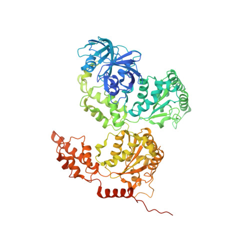
    p97 belongs to the superfamily of AAA+ ATPases and is characterized by a tandem AAA module, an N-terminal domain involved in substrate and cofactor interactions, and a functionally important unstructured C-terminal tail. The ATPase activity is controlled by an intradomain communication within the same protomer and an interdomain communication between neighboring protomers. Here, we present for the first time crystal structures in which the physiologically relevant p97 hexamer constitutes the content of the asymmetric unit, namely in the apo state without nucleotide in either the D1 or D2 module and in the pre-activated state with ATPγS bound to both modules. The structures provide new mechanistic insights into the interdomain communication mediated by conformational changes of the C terminus as well as an intersubunit signaling network, which couples the nucleotide state to the conformation of the central putative substrate binding pore.


  • Organizational Affiliation

    Rudolf Virchow Center for Experimental Biomedicine, University of Würzburg, Josef-Schneider-Strasse 2, 97080 Würzburg, Germany. Electronic address: petra.haenzelmann@virchow.uni-wuerzburg.de.


Macromolecules
Find similar proteins by:  (by identity cutoff)  |  3D Structure
Entity ID: 1
MoleculeChains Sequence LengthOrganismDetailsImage
Transitional endoplasmic reticulum ATPase
A, B, C, D, E
A, B, C, D, E, F
805Homo sapiensMutation(s): 4 
Gene Names: VCP
EC: 3.6.4.6
UniProt & NIH Common Fund Data Resources
Find proteins for P55072 (Homo sapiens)
Explore P55072 
Go to UniProtKB:  P55072
PHAROS:  P55072
GTEx:  ENSG00000165280 
Entity Groups  
Sequence Clusters30% Identity50% Identity70% Identity90% Identity95% Identity100% Identity
UniProt GroupP55072
Sequence Annotations
Expand
  • Reference Sequence
Small Molecules
Ligands 1 Unique
IDChains Name / Formula / InChI Key2D Diagram3D Interactions
SO4
Query on SO4

Download Ideal Coordinates CCD File 
G [auth A]
H [auth A]
I [auth B]
J [auth B]
K [auth C]
G [auth A],
H [auth A],
I [auth B],
J [auth B],
K [auth C],
L [auth C],
M [auth D],
N [auth D],
O [auth E],
P [auth E],
Q [auth F],
R [auth F]
SULFATE ION
O4 S
QAOWNCQODCNURD-UHFFFAOYSA-L
Experimental Data & Validation

Experimental Data

  • Method: X-RAY DIFFRACTION
  • Resolution: 4.20 Å
  • R-Value Free: 0.302 
  • R-Value Work: 0.237 
  • R-Value Observed: 0.240 
  • Space Group: P 21 21 21
Unit Cell:
Length ( Å )Angle ( ˚ )
a = 137.665α = 90
b = 165.78β = 90
c = 243.992γ = 90
Software Package:
Software NamePurpose
PHENIXrefinement
Aimlessdata scaling
MxCuBEdata collection
PDB_EXTRACTdata extraction
PHASERphasing

Structure Validation

View Full Validation Report


Entry History 

Deposition Data

Revision History  (Full details and data files)

  • Version 1.0: 2016-01-13
    Type: Initial release
  • Version 1.1: 2016-01-20
    Changes: Database references
  • Version 1.2: 2024-01-10
    Changes: Data collection, Database references, Refinement description