Characterization of a Poly(Butylene Adipate-Co-Terephthalate)-Hydrolyzing Lipase from Pelosinus Fermentans.
Biundo, A., Hromic, A., Pavkov-Keller, T., Gruber, K., Quartinello, F., Haernvall, K., Perz, V., Arrell, M.S., Zinn, M., Ribitsch, D., Guebitz, G.M.(2016) Appl Microbiol Biotechnol 100: 1753
- PubMed: 26490551
- DOI: https://doi.org/10.1007/s00253-015-7031-1
- Primary Citation of Related Structures:
5AH0 - PubMed Abstract:
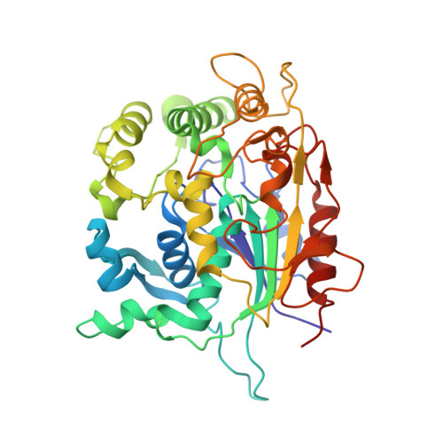
Certain α/β hydrolases have the ability to hydrolyze synthetic polyesters. While their partial hydrolysis has a potential for surface functionalization, complete hydrolysis allows recycling of valuable building blocks. Although knowledge about biodegradation of these materials is important regarding their fate in the environment, it is currently limited to aerobic organisms. A lipase from the anaerobic groundwater organism Pelosinus fermentans DSM 17108(PfL1) was cloned and expressed in Escherichia coli BL21-Gold (DE3) and purified from the cell extract. Biochemical characterization with small substrates showed thermoalkalophilic properties (Topt=50 °C, pHopt=7.5) and higher activity towards para-nitrophenyl octanoate (12.7 U mg(-1)) compared to longer and shorter chain lengths (C14 0.7 U mg(-1) and C2 4.3 U mg(-1), respectively). Crystallization and determination of the 3-D structure displayed the presence of a lid structure and a zinc ion surrounded by an extra domain. These properties classify the enzyme into the I.5 lipase family. PfL1 is able to hydrolyze poly(1,4-butylene adipate-co-terephthalate) (PBAT) polymeric substrates. The hydrolysis of PBAT showed the release of small building blocks as detected by liquid chromatography mass spectrometry (LC-MS). Protein dynamics seem to be involved with lid opening for the hydrolysis of PBAT by PfL1.
Organizational Affiliation:
Institute of Environmental Biotechnology, University of Natural Resources and Life Science (BOKU), Konrad Lorenz Strasse 22, 3430, Tulln an der Donau, Austria.