Structural and Dynamics Studies of a Truncated Variant of Ci Repressor from Bacteriophage Tp901-1.
Rasmussen, K.K., Frandsen, K.E.H., Boeri Erba, E., Pedersen, M., Varming, A.K., Hammer, K., Kilstrup, M., Thulstrup, P.W., Blackledge, M., Jensen, M.R., Lo Leggio, L.(2016) Sci Rep 6: 29574
- PubMed: 27403839
- DOI: https://doi.org/10.1038/srep29574
- Primary Citation of Related Structures:
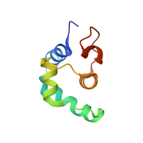
5A7L - PubMed Abstract:
The CI repressor from the temperate bacteriophage TP901-1 consists of two folded domains, an N-terminal helix-turn-helix DNA-binding domain (NTD) and a C-terminal oligomerization domain (CTD), which we here suggest to be further divided into CTD1 and CTD2. Full-length CI is a hexameric protein, whereas a truncated version, CI∆58, forms dimers. We identify the dimerization region of CI∆58 as CTD1 and determine its secondary structure to be helical both within the context of CI∆58 and in isolation. To our knowledge this is the first time that a helical dimerization domain has been found in a phage repressor. We also precisely determine the length of the flexible linker connecting the NTD to the CTD. Using electrophoretic mobility shift assays and native mass spectrometry, we show that CI∆58 interacts with the OL operator site as one dimer bound to both half-sites, and with much higher affinity than the isolated NTD domain thus demonstrating cooperativity between the two DNA binding domains. Finally, using small angle X-ray scattering data and state-of-the-art ensemble selection techniques, we delineate the conformational space sampled by CI∆58 in solution, and we discuss the possible role that the dynamics play in CI-repressor function.
Organizational Affiliation:
Department of Chemistry, University of Copenhagen, Universitetsparken 5, Copenhagen, Denmark.