5A4Q

DYRK1A IN COMPLEX WITH CHLORO BENZOTHIAZOLE FRAGMENT


Experimental Data Snapshot

  • Method: X-RAY DIFFRACTION
  • Resolution: 2.37 Å
  • R-Value Free: 0.243 
  • R-Value Work: 0.207 
  • R-Value Observed: 0.208 

wwPDB Validation   3D Report Full Report


Ligand Structure Quality Assessment 


This is version 1.6 of the entry. See complete history


Literature

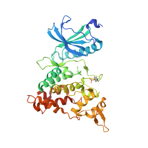
Probing the ATP-Binding Pocket of Protein Kinase Dyrk1A with Benzothiazole Fragment Molecules

Rothweiler, U.Stensen, W.Brandsdal, B.O.Isaksson, J.Leeson, F.A.Eugh, R.A.Mjoen Svendsen, J.S.

(2016) J Med Chem 59: 9814

  • DOI: https://doi.org/10.1021/acs.jmedchem.6b01086
  • Primary Citation of Related Structures:  
    5A3X, 5A4E, 5A4L, 5A4Q, 5A4T, 5A54

  • PubMed Abstract: 

    DYRK1A has emerged as a potential target for therapies of Alzheimer's disease using small molecules. On the basis of the observation of selective DYRK1A inhibition by firefly d-luciferin, we have explored static and dynamic structural properties of fragment sized variants of the benzothiazole scaffold with respect to DYRK1A using X-ray crystallography and NMR techniques. The compounds have excellent ligand efficiencies and show a remarkable diversity of binding modes in dynamic equilibrium. Binding geometries are determined in part by interactions often considered "weak", including "orthogonal multipolar" types represented by, for example, F-CO, sulfur-aromatic, and halogen-aromatic interactions, together with hydrogen bonds that are modulated by variation of electron withdrawing groups. These studies show how the benzothiazole scaffold is highly promising for the development of therapeutic DYRK1A inhibitors. In addition, the subtleties of the binding interactions, including dynamics, show how full structural studies are required to fully interpret the essential physical determinants of binding.


  • Organizational Affiliation

    The Norwegian Structural Biology Centre, Department of Chemistry, UiT The Arctic University of Norway , N-9037 Tromsø, Norway.


Macromolecules
Find similar proteins by:  (by identity cutoff)  |  3D Structure
Entity ID: 1
MoleculeChains Sequence LengthOrganismDetailsImage
DUAL SPECIFICITY TYROSINE-PHOSPHORYLATION-REGULATED KINASE 1A
A, B, C, D
368Homo sapiensMutation(s): 0 
EC: 2.7.12.1
UniProt & NIH Common Fund Data Resources
Find proteins for Q13627 (Homo sapiens)
Explore Q13627 
Go to UniProtKB:  Q13627
PHAROS:  Q13627
GTEx:  ENSG00000157540 
Entity Groups  
Sequence Clusters30% Identity50% Identity70% Identity90% Identity95% Identity100% Identity
UniProt GroupQ13627
Sequence Annotations
Expand
  • Reference Sequence
Small Molecules
Modified Residues  1 Unique
IDChains TypeFormula2D DiagramParent
PTR
Query on PTR
A, B, C, D
L-PEPTIDE LINKINGC9 H12 N O6 PTYR
Binding Affinity Annotations 
IDSourceBinding Affinity
Y3L Binding MOAD:  5A4Q IC50: 2900 (nM) from 1 assay(s)
Experimental Data & Validation

Experimental Data

  • Method: X-RAY DIFFRACTION
  • Resolution: 2.37 Å
  • R-Value Free: 0.243 
  • R-Value Work: 0.207 
  • R-Value Observed: 0.208 
  • Space Group: P 21 21 21
Unit Cell:
Length ( Å )Angle ( ˚ )
a = 88.039α = 90
b = 88.938β = 90
c = 229.759γ = 90
Software Package:
Software NamePurpose
REFMACrefinement
XDSdata reduction
XDSdata scaling
PHASERphasing

Structure Validation

View Full Validation Report


Ligand Structure Quality Assessment 


Entry History 

Deposition Data

Revision History  (Full details and data files)

  • Version 1.0: 2016-06-29
    Type: Initial release
  • Version 1.1: 2016-11-23
    Changes: Database references
  • Version 1.2: 2016-12-14
    Changes: Database references
  • Version 1.3: 2016-12-28
    Changes: Database references
  • Version 1.4: 2019-01-30
    Changes: Data collection, Experimental preparation, Other
  • Version 1.5: 2019-02-06
    Changes: Data collection, Experimental preparation
  • Version 1.6: 2024-01-10
    Changes: Data collection, Database references, Derived calculations, Other, Refinement description