4ZVZ

Co-crystal structures of PP5 in complex with 5-methyl-7-oxabicyclo[2.2.1]heptane-2,3-dicarboxylic acid


Experimental Data Snapshot

  • Method: X-RAY DIFFRACTION
  • Resolution: 2.00 Å
  • R-Value Free: 0.235 
  • R-Value Work: 0.204 
  • R-Value Observed: 0.206 

wwPDB Validation   3D Report Full Report


Ligand Structure Quality Assessment 


This is version 1.2 of the entry. See complete history


Literature

Crystal structures and mutagenesis of PPP-family ser/thr protein phosphatases elucidate the selectivity of cantharidin and novel norcantharidin-based inhibitors of PP5C.

Chattopadhyay, D.Swingle, M.R.Salter, E.A.Wood, E.D'Arcy, B.Zivanov, C.Abney, K.Musiyenko, A.Rusin, S.F.Kettenbach, A.Yet, L.Schroeder, C.E.Golden, J.E.Dunham, W.H.Gingras, A.C.Banerjee, S.Forbes, D.Wierzbicki, A.Honkanen, R.E.

(2016) Biochem Pharmacol 109: 14-26

  • DOI: https://doi.org/10.1016/j.bcp.2016.03.011
  • Primary Citation of Related Structures:  
    4ZVZ, 4ZX2

  • PubMed Abstract: 

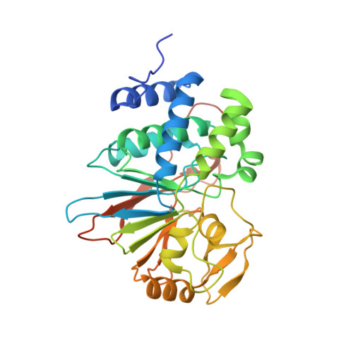
    Cantharidin is a natural toxin and an active constituent in a traditional Chinese medicine used to treat tumors. Cantharidin acts as a semi-selective inhibitor of PPP-family ser/thr protein phosphatases. Despite sharing a common catalytic mechanism and marked structural similarity with PP1C, PP2AC and PP5C, human PP4C was found to be insensitive to the inhibitory activity of cantharidin. To explore the molecular basis for this selectivity, we synthesized and tested novel C5/C6-derivatives designed from quantum-based modeling of the interactions revealed in the co-crystal structures of PP5C in complex with cantharidin. Structure-activity relationship studies and analysis of high-resolution (1.25Å) PP5C-inhibitor co-crystal structures reveal close contacts between the inhibitor bridgehead oxygen and both a catalytic metal ion and a non-catalytic phenylalanine residue, the latter of which is substituted by tryptophan in PP4C. Quantum chemistry calculations predicted that steric clashes with the bulkier tryptophan side chain in PP4C would force all cantharidin-based inhibitors into an unfavorable binding mode, disrupting the strong coordination of active site metal ions observed in the PP5C co-crystal structures, thereby rendering PP4C insensitive to the inhibitors. This prediction was confirmed by inhibition studies employing native human PP4C. Mutation of PP5C (F446W) and PP1C (F257W), to mimic the PP4C active site, resulted in markedly suppressed sensitivity to cantharidin. These observations provide insight into the structural basis for the natural selectivity of cantharidin and provide an avenue for PP4C deselection. The novel crystal structures also provide insight into interactions that provide increased selectivity of the C5/C6 modifications for PP5C versus other PPP-family phosphatases.


  • Organizational Affiliation

    Department of Medicine, University of Alabama Birmingham, Birmingham, AL 35294, USA.


Macromolecules
Find similar proteins by:  (by identity cutoff)  |  3D Structure
Entity ID: 1
MoleculeChains Sequence LengthOrganismDetailsImage
Serine/threonine-protein phosphatase 5
A, B, C, D
333Homo sapiensMutation(s): 0 
Gene Names: PPP5CPPP5
EC: 3.1.3.16
UniProt & NIH Common Fund Data Resources
Find proteins for P53041 (Homo sapiens)
Explore P53041 
Go to UniProtKB:  P53041
PHAROS:  P53041
GTEx:  ENSG00000011485 
Entity Groups  
Sequence Clusters30% Identity50% Identity70% Identity90% Identity95% Identity100% Identity
UniProt GroupP53041
Sequence Annotations
Expand
  • Reference Sequence
Small Molecules
Ligands 4 Unique
IDChains Name / Formula / InChI Key2D Diagram3D Interactions
4TF
Query on 4TF

Download Ideal Coordinates CCD File 
G [auth A],
J [auth B],
M [auth C],
R [auth D]
(1R,2S,3R,4S,5S)-5-(propoxymethyl)-7-oxabicyclo[2.2.1]heptane-2,3-dicarboxylic acid
C12 H18 O6
KENGHAXMPKBHLS-CHHOWFRJSA-N
PEG
Query on PEG

Download Ideal Coordinates CCD File 
O [auth C],
S [auth D]
DI(HYDROXYETHYL)ETHER
C4 H10 O3
MTHSVFCYNBDYFN-UHFFFAOYSA-N
MN
Query on MN

Download Ideal Coordinates CCD File 
E [auth A]
F [auth A]
H [auth B]
I [auth B]
K [auth C]
E [auth A],
F [auth A],
H [auth B],
I [auth B],
K [auth C],
L [auth C],
P [auth D],
Q [auth D]
MANGANESE (II) ION
Mn
WAEMQWOKJMHJLA-UHFFFAOYSA-N
NA
Query on NA

Download Ideal Coordinates CCD File 
N [auth C]SODIUM ION
Na
FKNQFGJONOIPTF-UHFFFAOYSA-N
Experimental Data & Validation

Experimental Data

  • Method: X-RAY DIFFRACTION
  • Resolution: 2.00 Å
  • R-Value Free: 0.235 
  • R-Value Work: 0.204 
  • R-Value Observed: 0.206 
  • Space Group: P 1
Unit Cell:
Length ( Å )Angle ( ˚ )
a = 40.858α = 88.15
b = 80.52β = 87.33
c = 91.165γ = 86.71
Software Package:
Software NamePurpose
REFMACrefinement
Aimlessdata scaling
PHENIXphasing
PDB_EXTRACTdata extraction
XDSdata reduction

Structure Validation

View Full Validation Report


Ligand Structure Quality Assessment 


Entry History 

Deposition Data

Revision History  (Full details and data files)

  • Version 1.0: 2016-04-27
    Type: Initial release
  • Version 1.1: 2017-02-08
    Changes: Database references
  • Version 1.2: 2023-09-27
    Changes: Data collection, Database references, Derived calculations, Refinement description