4ZU4

X-ray structure of the 3,4-ketoisomerase domain of FdtD from Shewanella denitrificans


Experimental Data Snapshot

  • Method: X-RAY DIFFRACTION
  • Resolution: 1.70 Å
  • R-Value Free: 0.207 
  • R-Value Work: 0.171 
  • R-Value Observed: 0.173 

wwPDB Validation   3D Report Full Report


Ligand Structure Quality Assessment 


This is version 1.3 of the entry. See complete history


Literature

Bacterial Sugar 3,4-Ketoisomerases: Structural Insight into Product Stereochemistry.

Thoden, J.B.Vinogradov, E.Gilbert, M.Salinger, A.J.Holden, H.M.

(2015) Biochemistry 54: 4495-4506

  • DOI: https://doi.org/10.1021/acs.biochem.5b00541
  • Primary Citation of Related Structures:  
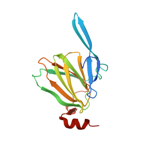
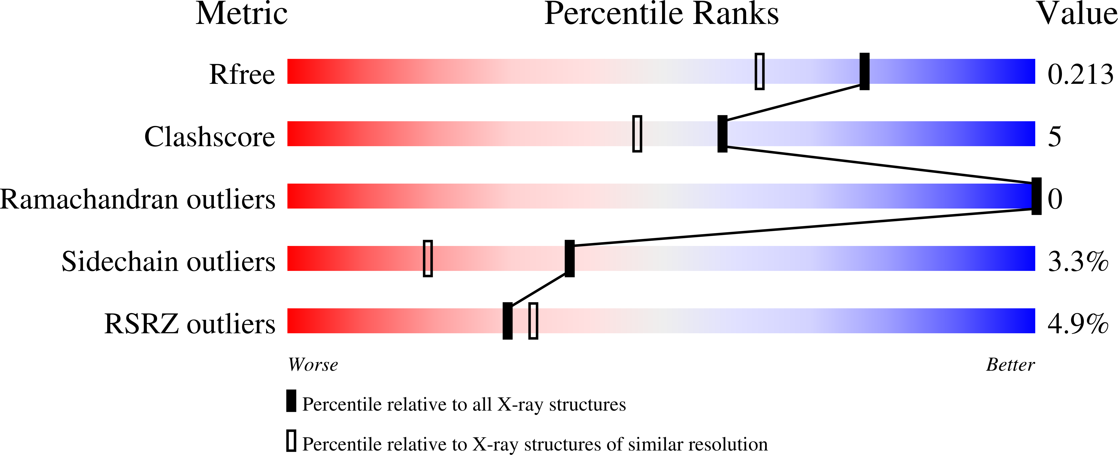
    4ZU4, 4ZU5, 4ZU7

  • PubMed Abstract: 

    3-Acetamido-3,6-dideoxy-d-galactose (Fuc3NAc) and 3-acetamido-3,6-dideoxy-d-glucose (Qui3NAc) are unusual sugars found on the lipopolysaccharides of Gram-negative bacteria and on the S-layers of Gram-positive bacteria. The 3,4-ketoisomerases, referred to as FdtA and QdtA, catalyze the third steps in the respective biosynthetic pathways for these sugars. Whereas both enzymes utilize the same substrate, the stereochemistries of their products are different. Specifically, the hydroxyl groups at the hexose C-4' positions assume the "galactose" and "glucose" configurations in the FdtA and QdtA products, respectively. In 2007 we reported the structure of the apoform of FdtA from Aneurinibacillus thermoaerophilus, which was followed in 2014 by the X-ray analysis of QdtA from Thermoanaerobacterium thermosaccharolyticum as a binary complex. Both of these enzymes belong to the cupin superfamily. Here we report a combined structural and enzymological study to explore the manner in which these enzymes control the stereochemistry of their products. Various site-directed mutant proteins of each enzyme were constructed, and their dTDP-sugar products were analyzed by NMR spectroscopy. In addition, the kinetic parameters for these protein variants were measured, and the structure of one, namely, the QdtA Y17R/R97H double mutant form, was determined to 2.3-Å resolution. Finally, in an attempt to obtain a model of FdtA with a bound dTDP-linked sugar, the 3,4-ketoisomerase domain of a bifunctional enzyme from Shewanella denitrificans was cloned, purified, and crystallized in the presence of a dTDP-linked sugar analogue. Taken together, the results from this investigation demonstrate that it is possible to convert a "galacto" enzyme into a "gluco" enzyme and vice versa.


  • Organizational Affiliation

    †Department of Biochemistry, University of Wisconsin, Madison, Wisconsin 53706, United States.


Macromolecules
Find similar proteins by:  (by identity cutoff)  |  3D Structure
Entity ID: 1
MoleculeChains Sequence LengthOrganismDetailsImage
WxcM-like protein
A, B, C, D, E
A, B, C, D, E, F
148Shewanella denitrificans OS217Mutation(s): 0 
Gene Names: Sden_2659
UniProt
Find proteins for Q12KT8 (Shewanella denitrificans (strain OS217 / ATCC BAA-1090 / DSM 15013))
Explore Q12KT8 
Go to UniProtKB:  Q12KT8
Entity Groups  
Sequence Clusters30% Identity50% Identity70% Identity90% Identity95% Identity100% Identity
UniProt GroupQ12KT8
Sequence Annotations
Expand
  • Reference Sequence
Small Molecules
Ligands 5 Unique
IDChains Name / Formula / InChI Key2D Diagram3D Interactions
4TG
Query on 4TG

Download Ideal Coordinates CCD File 
G [auth A],
L [auth B],
O [auth C]
dTDP-4,6-dideoxy-4-formamido-glucose
C17 H27 N3 O15 P2
QULUVRDLMBXPHO-GJSHGOAISA-N
PO4
Query on PO4

Download Ideal Coordinates CCD File 
V [auth E]PHOSPHATE ION
O4 P
NBIIXXVUZAFLBC-UHFFFAOYSA-K
EDO
Query on EDO

Download Ideal Coordinates CCD File 
I [auth A]
J [auth A]
N [auth B]
T [auth E]
U [auth E]
I [auth A],
J [auth A],
N [auth B],
T [auth E],
U [auth E],
X [auth F]
1,2-ETHANEDIOL
C2 H6 O2
LYCAIKOWRPUZTN-UHFFFAOYSA-N
CL
Query on CL

Download Ideal Coordinates CCD File 
H [auth A]
M [auth B]
Q [auth D]
R [auth D]
S [auth E]
H [auth A],
M [auth B],
Q [auth D],
R [auth D],
S [auth E],
W [auth F]
CHLORIDE ION
Cl
VEXZGXHMUGYJMC-UHFFFAOYSA-M
NA
Query on NA

Download Ideal Coordinates CCD File 
K [auth A],
P [auth C]
SODIUM ION
Na
FKNQFGJONOIPTF-UHFFFAOYSA-N
Experimental Data & Validation

Experimental Data

  • Method: X-RAY DIFFRACTION
  • Resolution: 1.70 Å
  • R-Value Free: 0.207 
  • R-Value Work: 0.171 
  • R-Value Observed: 0.173 
  • Space Group: P 1 21 1
Unit Cell:
Length ( Å )Angle ( ˚ )
a = 85.405α = 90
b = 64.579β = 111.39
c = 112.968γ = 90
Software Package:
Software NamePurpose
REFMACrefinement
SAINTdata reduction
SADABSdata scaling
PHASERphasing

Structure Validation

View Full Validation Report


Ligand Structure Quality Assessment 


Entry History 

Deposition Data

Revision History  (Full details and data files)

  • Version 1.0: 2015-07-15
    Type: Initial release
  • Version 1.1: 2015-07-22
    Changes: Database references
  • Version 1.2: 2015-08-05
    Changes: Database references
  • Version 1.3: 2023-09-27
    Changes: Data collection, Database references, Derived calculations, Refinement description