4ZTC

PglE Aminotransferase in complex with External Aldimine, Mutant K184A


Experimental Data Snapshot

  • Method: X-RAY DIFFRACTION
  • Resolution: 2.00 Å
  • R-Value Free: 0.254 
  • R-Value Work: 0.188 
  • R-Value Observed: 0.192 

wwPDB Validation   3D Report Full Report


Ligand Structure Quality Assessment 


This is version 1.2 of the entry. See complete history


Literature

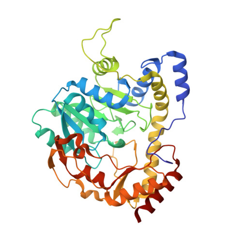
Structure of the external aldimine form of PglE, an aminotransferase required for N,N'-diacetylbacillosamine biosynthesis.

Riegert, A.S.Young, N.M.Watson, D.C.Thoden, J.B.Holden, H.M.

(2015) Protein Sci 24: 1609-1616

  • DOI: https://doi.org/10.1002/pro.2745
  • Primary Citation of Related Structures:  
    4ZTC

  • PubMed Abstract: 

    N,N'-diacetylbacillosamine is a novel sugar that plays a key role in bacterial glycosylation. Three enzymes are required for its biosynthesis in Campylobacter jejuni starting from UDP-GlcNAc. The focus of this investigation, PglE, catalyzes the second step in the pathway. It is a PLP-dependent aminotransferase that converts UDP-2-acetamido-4-keto-2,4,6-trideoxy-d-glucose to UDP-2-acetamido-4-amino-2,4,6-trideoxy-d-glucose. For this investigation, the structure of PglE in complex with an external aldimine was determined to a nominal resolution of 2.0 Å. A comparison of its structure with those of other sugar aminotransferases reveals a remarkable difference in the manner by which PglE accommodates its nucleotide-linked sugar substrate.


  • Organizational Affiliation

    Department of Biochemistry, University of Wisconsin, Madison, Wisconsin, 53706.


Macromolecules
Find similar proteins by:  (by identity cutoff)  |  3D Structure
Entity ID: 1
MoleculeChains Sequence LengthOrganismDetailsImage
Aminotransferase homolog386Campylobacter jejuniMutation(s): 1 
UniProt
Find proteins for Q0P9D3 (Campylobacter jejuni subsp. jejuni serotype O:2 (strain ATCC 700819 / NCTC 11168))
Explore Q0P9D3 
Go to UniProtKB:  Q0P9D3
Entity Groups  
Sequence Clusters30% Identity50% Identity70% Identity90% Identity95% Identity100% Identity
UniProt GroupQ0P9D3
Sequence Annotations
Expand
  • Reference Sequence
Small Molecules
Ligands 1 Unique
IDChains Name / Formula / InChI Key2D Diagram3D Interactions
4RA
Query on 4RA

Download Ideal Coordinates CCD File 
B [auth A][(2R,3R,4R,5S,6R)-3-acetamido-6-methyl-5-[(E)-[2-methyl-3-oxidanyl-5-(phosphonooxymethyl)pyridin-4-yl]methylideneamino]-4-oxidanyl-oxan-2-yl] [[(2R,3S,4R,5R)-5-[2,4-bis(oxidanylidene)pyrimidin-1-yl]-3,4-bis(oxidanyl)oxolan-2-yl]methoxy-oxidanyl-phosphoryl] hydrogen phosphate
C25 H36 N5 O20 P3
BKHPSAUTHKIVAK-OTBUILFPSA-N
Experimental Data & Validation

Experimental Data

  • Method: X-RAY DIFFRACTION
  • Resolution: 2.00 Å
  • R-Value Free: 0.254 
  • R-Value Work: 0.188 
  • R-Value Observed: 0.192 
  • Space Group: P 41 21 2
Unit Cell:
Length ( Å )Angle ( ˚ )
a = 66.161α = 90
b = 66.161β = 90
c = 169.013γ = 90
Software Package:
Software NamePurpose
REFMACrefinement
SAINTdata reduction
SADABSdata scaling
PHASERphasing

Structure Validation

View Full Validation Report


Ligand Structure Quality Assessment 


Entry History 

Deposition Data

Revision History  (Full details and data files)

  • Version 1.0: 2015-07-29
    Type: Initial release
  • Version 1.1: 2015-10-14
    Changes: Database references
  • Version 1.2: 2023-09-27
    Changes: Data collection, Database references, Derived calculations, Refinement description