4ZCV

Structure of calcium-bound regulatory domain of the human ATP-Mg/Pi carrier in the P212121 form


Experimental Data Snapshot

  • Method: X-RAY DIFFRACTION
  • Resolution: 2.80 Å
  • R-Value Free: 0.259 
  • R-Value Work: 0.211 
  • R-Value Observed: 0.213 

wwPDB Validation   3D Report Full Report


This is version 1.3 of the entry. See complete history


Literature

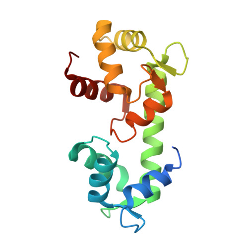
Calcium-induced conformational changes in the regulatory domain of the human mitochondrial ATP-Mg/Pi carrier.

Harborne, S.P.Ruprecht, J.J.Kunji, E.R.

(2015) Biochim Biophys Acta 1847: 1245-1253

  • DOI: https://doi.org/10.1016/j.bbabio.2015.07.002
  • Primary Citation of Related Structures:  
    4ZCU, 4ZCV

  • PubMed Abstract: 

    The mitochondrial ATP-Mg/Pi carrier imports adenine nucleotides from the cytosol into the mitochondrial matrix and exports phosphate. The carrier is regulated by the concentration of cytosolic calcium, altering the size of the adenine nucleotide pool in the mitochondrial matrix in response to energetic demands. The protein consists of three domains; (i) the N-terminal regulatory domain, which is formed of two pairs of fused calcium-binding EF-hands, (ii) the C-terminal mitochondrial carrier domain, which is involved in transport, and (iii) a linker region with an amphipathic α-helix of unknown function. The mechanism by which calcium binding to the regulatory domain modulates substrate transport in the carrier domain has not been resolved. Here, we present two new crystal structures of the regulatory domain of the human isoform 1. Careful analysis by SEC confirmed that although the regulatory domain crystallised as dimers, full-length ATP-Mg/Pi carrier is monomeric. Therefore, the ATP-Mg/Pi carrier must have a different mechanism of calcium regulation than the architecturally related aspartate/glutamate carrier, which is dimeric. The structure showed that an amphipathic α-helix is bound to the regulatory domain in a hydrophobic cleft of EF-hand 3/4. Detailed bioinformatics analyses of different EF-hand states indicate that upon release of calcium, EF-hands close, meaning that the regulatory domain would release the amphipathic α-helix. We propose a mechanism for ATP-Mg/Pi carriers in which the amphipathic α-helix becomes mobile upon release of calcium and could block the transport of substrates across the mitochondrial inner membrane.


  • Organizational Affiliation

    The Medical Research Council, Mitochondrial Biology Unit, Cambridge Biomedical Campus, Wellcome Trust/MRC Building, Hills Road, Cambridge CB2 0XY, UK.


Macromolecules
Find similar proteins by:  (by identity cutoff)  |  3D Structure
Entity ID: 1
MoleculeChains Sequence LengthOrganismDetailsImage
Calcium-binding mitochondrial carrier protein SCaMC-1
A, B, C, D
165Homo sapiensMutation(s): 0 
Gene Names: SLC25A24APC1MCSC1SCAMC1
UniProt & NIH Common Fund Data Resources
Find proteins for Q6NUK1 (Homo sapiens)
Explore Q6NUK1 
Go to UniProtKB:  Q6NUK1
PHAROS:  Q6NUK1
GTEx:  ENSG00000085491 
Entity Groups  
Sequence Clusters30% Identity50% Identity70% Identity90% Identity95% Identity100% Identity
UniProt GroupQ6NUK1
Sequence Annotations
Expand
  • Reference Sequence
Small Molecules
Ligands 3 Unique
IDChains Name / Formula / InChI Key2D Diagram3D Interactions
SO4
Query on SO4

Download Ideal Coordinates CCD File 
H [auth A]
I [auth A]
K [auth A]
O [auth B]
U [auth C]
H [auth A],
I [auth A],
K [auth A],
O [auth B],
U [auth C],
Z [auth D]
SULFATE ION
O4 S
QAOWNCQODCNURD-UHFFFAOYSA-L
GOL
Query on GOL

Download Ideal Coordinates CCD File 
J [auth A],
P [auth B],
V [auth C]
GLYCEROL
C3 H8 O3
PEDCQBHIVMGVHV-UHFFFAOYSA-N
CA
Query on CA

Download Ideal Coordinates CCD File 
E [auth A]
F [auth A]
G [auth A]
L [auth B]
M [auth B]
E [auth A],
F [auth A],
G [auth A],
L [auth B],
M [auth B],
N [auth B],
Q [auth C],
R [auth C],
S [auth C],
T [auth C],
W [auth D],
X [auth D],
Y [auth D]
CALCIUM ION
Ca
BHPQYMZQTOCNFJ-UHFFFAOYSA-N
Experimental Data & Validation

Experimental Data

  • Method: X-RAY DIFFRACTION
  • Resolution: 2.80 Å
  • R-Value Free: 0.259 
  • R-Value Work: 0.211 
  • R-Value Observed: 0.213 
  • Space Group: P 21 21 21
Unit Cell:
Length ( Å )Angle ( ˚ )
a = 75.92α = 90
b = 77.01β = 90
c = 119.56γ = 90
Software Package:
Software NamePurpose
PHENIXrefinement
iMOSFLMdata reduction
Aimlessdata scaling
PHASERphasing
PDB_EXTRACTdata extraction

Structure Validation

View Full Validation Report


Entry History & Funding Information

Deposition Data


Funding OrganizationLocationGrant Number
Medical Research Council (United Kingdom)United Kingdom--

Revision History  (Full details and data files)

  • Version 1.0: 2015-07-22
    Type: Initial release
  • Version 1.1: 2015-07-29
    Changes: Database references
  • Version 1.2: 2017-09-13
    Changes: Author supporting evidence
  • Version 1.3: 2024-01-10
    Changes: Data collection, Database references, Derived calculations, Refinement description