


Crystal structure of PfaD from Shewanella oneidensis in complex with NAD+ determined by in-situ diffraction.
Baker, G.E., Race, P.R.To be published.
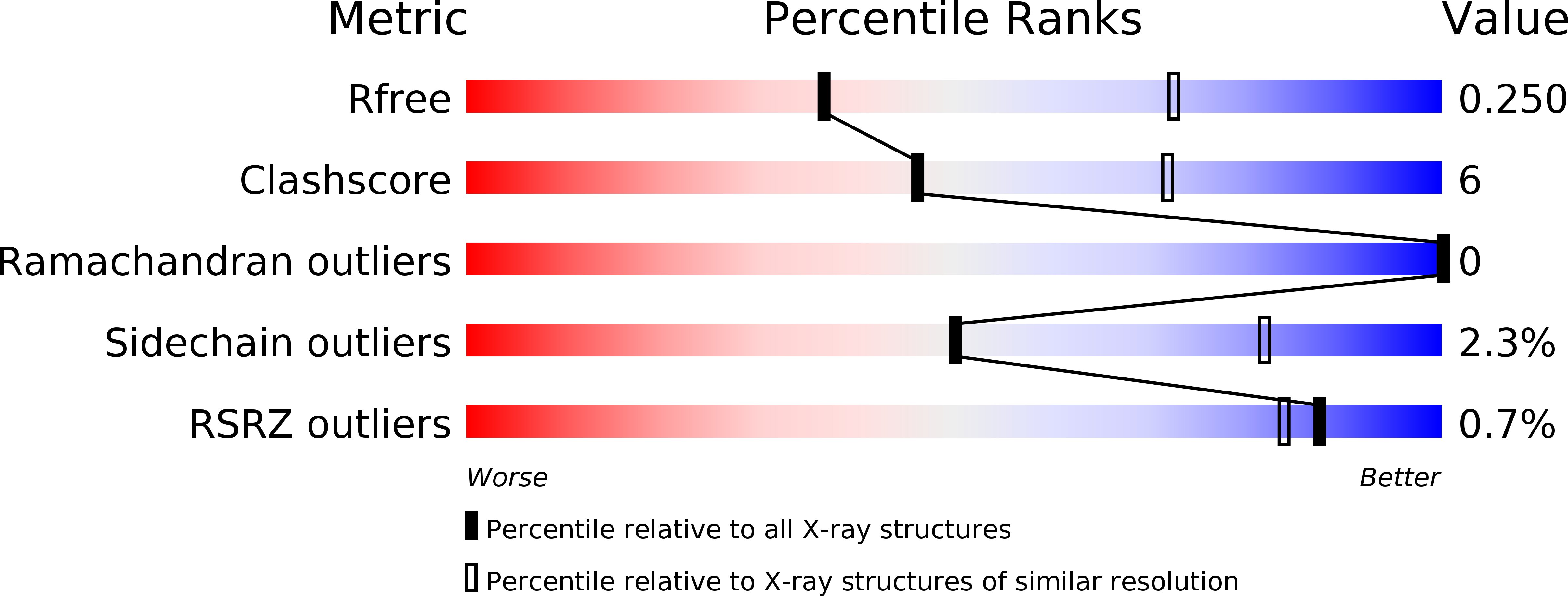
Experimental Data Snapshot
Entity ID: 1 | |||||
|---|---|---|---|---|---|
| Molecule | Chains | Sequence Length | Organism | Details | Image |
| Omega-3 polyunsaturated fatty acid synthase subunit PfaD | 547 | Shewanella oneidensis MR-1 | Mutation(s): 0 Gene Names: pfaD, SO_1597 | 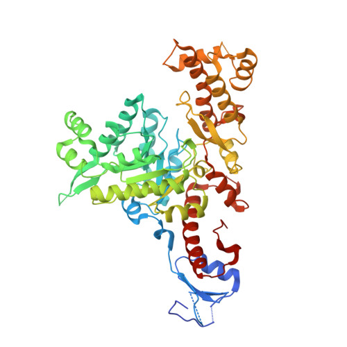 | |
UniProt | |||||
Find proteins for Q8EGK4 (Shewanella oneidensis (strain MR-1)) Explore Q8EGK4 Go to UniProtKB: Q8EGK4 | |||||
Entity Groups | |||||
| Sequence Clusters | 30% Identity50% Identity70% Identity90% Identity95% Identity100% Identity | ||||
| UniProt Group | Q8EGK4 | ||||
Sequence AnnotationsExpand | |||||
| |||||
| Ligands 2 Unique | |||||
|---|---|---|---|---|---|
| ID | Chains | Name / Formula / InChI Key | 2D Diagram | 3D Interactions | |
| NAD Query on NAD | C [auth A], E [auth B] | NICOTINAMIDE-ADENINE-DINUCLEOTIDE C21 H27 N7 O14 P2 BAWFJGJZGIEFAR-NNYOXOHSSA-N |  | ||
| FMN Query on FMN | D [auth A], F [auth B] | FLAVIN MONONUCLEOTIDE C17 H21 N4 O9 P FVTCRASFADXXNN-SCRDCRAPSA-N |  | ||
| Length ( Å ) | Angle ( ˚ ) |
|---|---|
| a = 106.68 | α = 90 |
| b = 106.68 | β = 90 |
| c = 217.477 | γ = 90 |
| Software Name | Purpose |
|---|---|
| REFMAC | refinement |
| PHASER | phasing |
| Coot | model building |
| XDS | data reduction |
| Aimless | data scaling |
RCSB PDB (citation) is hosted by
RCSB PDB is a member of the

