4Z9A

Crystal structure of Low Molecular Weight Protein Tyrosine Phosphatase isoform A complexed with phenylmethanesulfonic acid


Experimental Data Snapshot

  • Method: X-RAY DIFFRACTION
  • Resolution: 2.10 Å
  • R-Value Free: 0.235 
  • R-Value Work: 0.179 
  • R-Value Observed: 0.183 

wwPDB Validation   3D Report Full Report


Ligand Structure Quality Assessment 


This is version 1.5 of the entry. See complete history


Literature

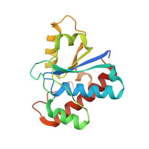
Crystal structures of the apo form and a complex of human LMW-PTP with a phosphonic acid provide new evidence of a secondary site potentially related to the anchorage of natural substrates.

Fonseca, E.M.Trivella, D.B.Scorsato, V.Dias, M.P.Bazzo, N.L.Mandapati, K.R.de Oliveira, F.L.Ferreira-Halder, C.V.Pilli, R.A.Miranda, P.C.Aparicio, R.

(2015) Bioorg Med Chem 23: 4462-4471

  • DOI: https://doi.org/10.1016/j.bmc.2015.06.017
  • Primary Citation of Related Structures:  
    4Z99, 4Z9A, 4Z9B

  • PubMed Abstract: 

    Low molecular weight protein tyrosine phosphatases (LMW-PTP, EC 3.1.3.48) are a family of single-domain enzymes with molecular weight up to 18 kDa, expressed in different tissues and considered attractive pharmacological targets for cancer chemotherapy. Despite this, few LMW-PTP inhibitors have been described to date, and the structural information on LMW-PTP druggable binding sites is scarce. In this study, a small series of phosphonic acids were designed based on a new crystallographic structure of LMW-PTP complexed with benzylsulfonic acid, determined at 2.1Å. In silico docking was used as a tool to interpret the structural and enzyme kinetics data, as well as to design new analogs. From the synthesized series, two compounds were found to act as competitive inhibitors, with inhibition constants of 0.124 and 0.047 mM. We also report the 2.4Å structure of another complex in which LMW-PTP is bound to benzylphosphonic acid, and a structure of apo LMW-PTP determined at 2.3Å resolution. Although no appreciable conformation changes were observed, in the latter structures, amino acid residues from an expression tag were found bound to a hydrophobic region at the protein surface. This regions is neighbored by positively charged residues, adjacent to the active site pocket, suggesting that this region might be not a mere artefact of crystal contacts but an indication of a possible anchoring region for the natural substrate-which is a phosphorylated protein.


  • Organizational Affiliation

    Laboratory of Structural Biology and Crystallography, Institute of Chemistry, University of Campinas, CP 6154, 13083-970, Campinas, SP, Brazil.


Macromolecules
Find similar proteins by:  (by identity cutoff)  |  3D Structure
Entity ID: 1
MoleculeChains Sequence LengthOrganismDetailsImage
Low molecular weight phosphotyrosine protein phosphatase164Homo sapiensMutation(s): 0 
Gene Names: ACP1
EC: 3.1.3.48 (PDB Primary Data), 3.1.3.2 (PDB Primary Data)
UniProt & NIH Common Fund Data Resources
Find proteins for P24666 (Homo sapiens)
Explore P24666 
Go to UniProtKB:  P24666
PHAROS:  P24666
GTEx:  ENSG00000143727 
Entity Groups  
Sequence Clusters30% Identity50% Identity70% Identity90% Identity95% Identity100% Identity
UniProt GroupP24666
Sequence Annotations
Expand
  • Reference Sequence
Experimental Data & Validation

Experimental Data

  • Method: X-RAY DIFFRACTION
  • Resolution: 2.10 Å
  • R-Value Free: 0.235 
  • R-Value Work: 0.179 
  • R-Value Observed: 0.183 
  • Space Group: P 21 21 21
Unit Cell:
Length ( Å )Angle ( ˚ )
a = 32.75α = 90
b = 55.23β = 90
c = 97.8γ = 90
Software Package:
Software NamePurpose
REFMACrefinement
MOSFLMdata reduction
SCALAdata scaling
PHASERphasing

Structure Validation

View Full Validation Report


Ligand Structure Quality Assessment 


Entry History & Funding Information

Deposition Data


Funding OrganizationLocationGrant Number
Sao Paulo Research Foundation (FAPESP)Brazil2009/51602-5
Sao Paulo Research Foundation (FAPESP)Brazil2010/17544-5
Sao Paulo Research Foundation (FAPESP)Brazil2011/15792-4
Sao Paulo Research Foundation (FAPESP)Brazil2011/03054-9

Revision History  (Full details and data files)

  • Version 1.0: 2015-07-15
    Type: Initial release
  • Version 1.1: 2015-08-05
    Changes: Database references
  • Version 1.2: 2018-01-17
    Changes: Author supporting evidence, Database references, Derived calculations
  • Version 1.3: 2019-04-17
    Changes: Author supporting evidence, Data collection
  • Version 1.4: 2020-01-01
    Changes: Author supporting evidence
  • Version 1.5: 2023-09-27
    Changes: Data collection, Database references, Refinement description