4Z0B

Crystal Structure of the Fab Fragment of Anti-ofloxacin Antibody and Exploration Its Receptor Binding Site


Experimental Data Snapshot

  • Method: X-RAY DIFFRACTION
  • Resolution: 3.20 Å
  • R-Value Free: 0.309 
  • R-Value Work: 0.276 
  • R-Value Observed: 0.278 

wwPDB Validation   3D Report Full Report


This is version 1.1 of the entry. See complete history


Literature

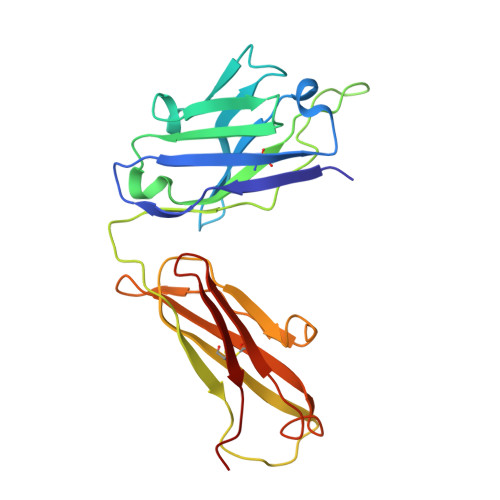
Crystal Structure of the Fab Fragment of an Anti-ofloxacin Antibody and Exploration of Its Specific Binding.

He, K.Du, X.Sheng, W.Zhou, X.Wang, J.Wang, S.

(2016) J Agric Food Chem 64: 2627-2634

  • DOI: https://doi.org/10.1021/acs.jafc.5b05882
  • Primary Citation of Related Structures:  
    4Z0B

  • PubMed Abstract: 

    The limited knowledge on the mechanism of interactions between small contaminants and the corresponding antibodies greatly inhibits the development of enzyme-linked immunosorbent assay methods. In this study, the crystal structure of a Fab fragment specific for ofloxacin was obtained. On the basis of the crystal characteristics, the modeling of the interactions between ofloxacin and the Fab revealed that TYR31 and HIS99 of the heavy chain and MET20 and GLN79 of the light chain formed a hydrophobic region and that SER52 and ALA97 of the heavy chain and TYR35 of the light chain formed a salt bridge and two hydrogen bonds for specific binding. The key roles of SER52 and ALA97 were further confirmed by site-directed mutation. A specificity analysis using 14 ofloxacin analogues indicates that the length of the bond formed between the piperazine ring and the antibody plays key roles in specific recognition. This work helps to clarify the mechanisms through which antibodies recognize small molecules and improve immune detection methods.


  • Organizational Affiliation

    Key Laboratory of Food Nutrition and Safety, Ministry of Education of China, Tianjin University of Science and Technology , Tianjin 300457, People's Republic of China.


Macromolecules
Find similar proteins by:  (by identity cutoff)  |  3D Structure
Entity ID: 1
MoleculeChains Sequence LengthOrganismDetailsImage
antibody heavy chainA [auth H]222Mus musculusMutation(s): 0 
Entity Groups  
Sequence Clusters30% Identity50% Identity70% Identity90% Identity95% Identity100% Identity
Sequence Annotations
Expand
  • Reference Sequence
Find similar proteins by:  (by identity cutoff)  |  3D Structure
Entity ID: 2
MoleculeChains Sequence LengthOrganismDetailsImage
antibody light chainB [auth L]212Mus musculusMutation(s): 0 
Entity Groups  
Sequence Clusters30% Identity50% Identity70% Identity90% Identity95% Identity100% Identity
Sequence Annotations
Expand
  • Reference Sequence
Small Molecules
Ligands 1 Unique
IDChains Name / Formula / InChI Key2D Diagram3D Interactions
PO4
Query on PO4

Download Ideal Coordinates CCD File 
C [auth H]PHOSPHATE ION
O4 P
NBIIXXVUZAFLBC-UHFFFAOYSA-K
Experimental Data & Validation

Experimental Data

  • Method: X-RAY DIFFRACTION
  • Resolution: 3.20 Å
  • R-Value Free: 0.309 
  • R-Value Work: 0.276 
  • R-Value Observed: 0.278 
  • Space Group: C 1 2 1
Unit Cell:
Length ( Å )Angle ( ˚ )
a = 62.828α = 90
b = 53.528β = 98.39
c = 129.103γ = 90
Software Package:
Software NamePurpose
HKL-2000data collection
HKL-2000data scaling
REFMACrefinement
PDB_EXTRACTdata extraction
HKL-2000data reduction
PHASERphasing

Structure Validation

View Full Validation Report


Entry History & Funding Information

Deposition Data


Funding OrganizationLocationGrant Number
National naural science foundation of chinaChina31225021

Revision History  (Full details and data files)

  • Version 1.0: 2016-04-27
    Type: Initial release
  • Version 1.1: 2023-11-08
    Changes: Data collection, Database references, Derived calculations, Refinement description