4YN8

Crystal Structure of Response Regulator ChrA in Heme-Sensing Two Component System


Experimental Data Snapshot

  • Method: X-RAY DIFFRACTION
  • Resolution: 1.80 Å
  • R-Value Free: 0.202 
  • R-Value Work: 0.176 
  • R-Value Observed: 0.177 

wwPDB Validation   3D Report Full Report


This is version 1.2 of the entry. See complete history


Literature

Structure of the response regulator ChrA in the haem-sensing two-component system of Corynebacterium diphtheriae.

Doi, A.Nakamura, H.Shiro, Y.Sugimoto, H.

(2015) Acta Crystallogr F Struct Biol Commun 71: 966-971

  • DOI: https://doi.org/10.1107/S2053230X15009838
  • Primary Citation of Related Structures:  
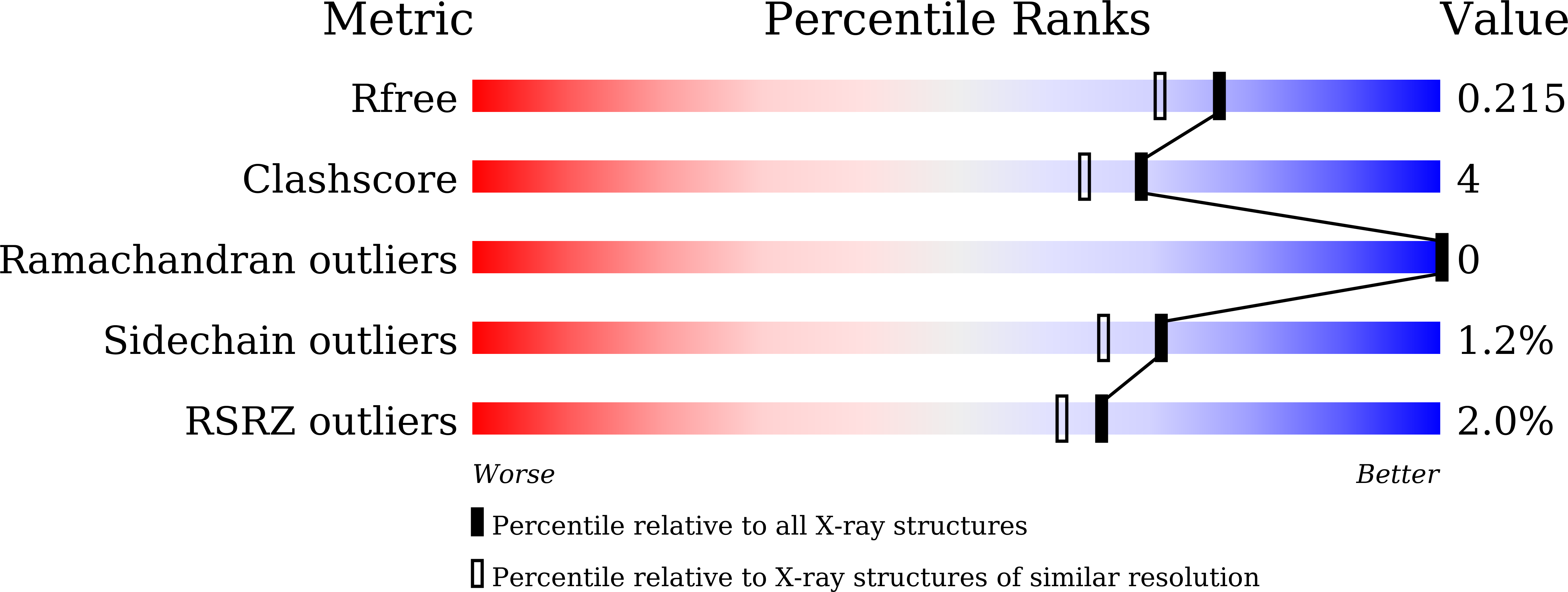
    4YN8

  • PubMed Abstract: 

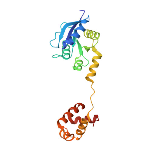
    ChrA is a response regulator (RR) in the two-component system involved in regulating the degradation and transport of haem (Fe-porphyrin) in the pathogen Corynebacterium diphtheriae. Here, the crystal structure of full-length ChrA is described at a resolution of 1.8 Å. ChrA consists of an N-terminal regulatory domain, a long linker region and a C-terminal DNA-binding domain. A structural comparison of ChrA with other RRs revealed substantial differences in the relative orientation of the two domains and the conformation of the linker region. The structural flexibility of the linker could be an important feature in rearrangement of the domain orientation to create a dimerization interface to bind DNA during haem-sensing signal transduction.


  • Organizational Affiliation

    RIKEN SPring-8 Center, 1-1-1 Kouto, Sayo, Hyogo 679-5148, Japan.


Macromolecules
Find similar proteins by:  (by identity cutoff)  |  3D Structure
Entity ID: 1
MoleculeChains Sequence LengthOrganismDetailsImage
Response regulator ChrA203Corynebacterium diphtheriaeMutation(s): 1 
Gene Names: chrA
UniProt
Find proteins for Q9RPE9 (Corynebacterium diphtheriae)
Explore Q9RPE9 
Go to UniProtKB:  Q9RPE9
Entity Groups  
Sequence Clusters30% Identity50% Identity70% Identity90% Identity95% Identity100% Identity
UniProt GroupQ9RPE9
Sequence Annotations
Expand
  • Reference Sequence
Small Molecules
Ligands 2 Unique
IDChains Name / Formula / InChI Key2D Diagram3D Interactions
SO4
Query on SO4

Download Ideal Coordinates CCD File 
B [auth A],
C [auth A],
D [auth A],
E [auth A]
SULFATE ION
O4 S
QAOWNCQODCNURD-UHFFFAOYSA-L
MG
Query on MG

Download Ideal Coordinates CCD File 
F [auth A]
G [auth A]
H [auth A]
I [auth A]
J [auth A]
F [auth A],
G [auth A],
H [auth A],
I [auth A],
J [auth A],
K [auth A],
L [auth A]
MAGNESIUM ION
Mg
JLVVSXFLKOJNIY-UHFFFAOYSA-N
Experimental Data & Validation

Experimental Data

  • Method: X-RAY DIFFRACTION
  • Resolution: 1.80 Å
  • R-Value Free: 0.202 
  • R-Value Work: 0.176 
  • R-Value Observed: 0.177 
  • Space Group: I 41 2 2
Unit Cell:
Length ( Å )Angle ( ˚ )
a = 84.88α = 90
b = 84.88β = 90
c = 169.3γ = 90
Software Package:
Software NamePurpose
HKL-2000data scaling
REFMACrefinement
PDB_EXTRACTdata extraction
BALBESphasing
Cootmodel building

Structure Validation

View Full Validation Report


Entry History & Funding Information

Deposition Data


Funding OrganizationLocationGrant Number
KAKENHIJapan24687015
KAKENHIJapan25121739
KAKENHIJapan23121531

Revision History  (Full details and data files)

  • Version 1.0: 2015-08-19
    Type: Initial release
  • Version 1.1: 2020-02-05
    Changes: Data collection, Derived calculations
  • Version 1.2: 2023-11-08
    Changes: Data collection, Database references, Derived calculations, Refinement description