4YKF

Crystal Structure of the Alkylhydroperoxide Reductase subunit F (AhpF) with NADH from Escherichia coli


Experimental Data Snapshot

  • Method: X-RAY DIFFRACTION
  • Resolution: 2.50 Å
  • R-Value Free: 0.288 
  • R-Value Work: 0.225 
  • R-Value Observed: 0.228 

wwPDB Validation   3D Report Full Report


Ligand Structure Quality Assessment 


This is version 1.1 of the entry. See complete history


Literature

Crystallographic and solution studies of NAD(+)- and NADH-bound alkylhydroperoxide reductase subunit F (AhpF) from Escherichia coli provide insight into sequential enzymatic steps

Kamariah, N.Manimekalai, M.S.S.Nartey, W.Eisenhaber, F.Eisenhaber, B.Gruber, G.

(2015) Biochim Biophys Acta 1847: 1139-1152

  • DOI: https://doi.org/10.1016/j.bbabio.2015.06.011
  • Primary Citation of Related Structures:  
    4YKF, 4YKG

  • PubMed Abstract: 

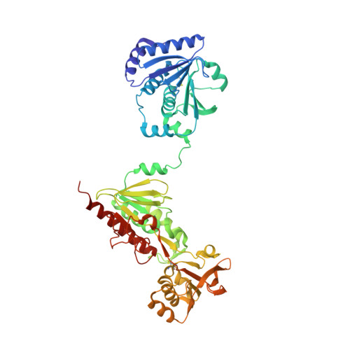
    Redox homeostasis is significant for the survival of pro- and eukaryotic cells and is crucial for defense against reactive oxygen species like superoxide and hydrogen peroxide. In Escherichia coli, the reduction of peroxides occurs via the redox active disulfide center of the alkyl hydroperoxide reductase C subunit (AhpC), whose reduced state becomes restored by AhpF. The 57kDa EcAhpF contains an N-terminal domain (NTD), which catalyzes the electron transfer from NADH via an FAD of the C-terminal domain into EcAhpC. The NTD is connected to the C-terminal domain via a linker. Here, the first crystal structure of E. coli AhpF bound with NADH and NAD(+) has been determined at 2.5Å and 2.4Å resolution, respectively. The NADH-bound form of EcAhpF reveals that the NADH-binding domain is required to alter its conformation to bring a bound NADH to the re-face of the isoalloxazine ring of the flavin, and thereby render the NADH-domain dithiol center accessible to the NTD disulfide center for electron transfer. The NAD(+)-bound form of EcAhpF shows conformational differences for the nicotinamide end moieties and its interacting residue M467, which is proposed to represent an intermediate product-release conformation. In addition, the structural alterations in EcAhpF due to NADH- and NAD(+)-binding in solution are shown by small angle X-ray scattering studies. The EcAhpF is revealed to adopt many intermediate conformations in solution to facilitate the electron transfer from the substrate NADH to the C-terminal domain, and subsequently to the NTD of EcAhpF for the final step of AhpC reduction.


  • Organizational Affiliation

    Bioinformatics Institute, Agency for Science, Technology and Research (A*STAR), 30 Biopolis Street, #07-01 Matrix, Singapore 138671, Republic of Singapore.


Macromolecules
Find similar proteins by:  (by identity cutoff)  |  3D Structure
Entity ID: 1
MoleculeChains Sequence LengthOrganismDetailsImage
Alkyl hydroperoxide reductase subunit F521Escherichia coli K-12Mutation(s): 0 
Gene Names: ahpFb0606JW0599
EC: 1.8.1
UniProt
Find proteins for P35340 (Escherichia coli (strain K12))
Explore P35340 
Go to UniProtKB:  P35340
Entity Groups  
Sequence Clusters30% Identity50% Identity70% Identity90% Identity95% Identity100% Identity
UniProt GroupP35340
Sequence Annotations
Expand
  • Reference Sequence
Small Molecules
Ligands 6 Unique
IDChains Name / Formula / InChI Key2D Diagram3D Interactions
FAD
Query on FAD

Download Ideal Coordinates CCD File 
B [auth A]FLAVIN-ADENINE DINUCLEOTIDE
C27 H33 N9 O15 P2
VWWQXMAJTJZDQX-UYBVJOGSSA-N
NAI
Query on NAI

Download Ideal Coordinates CCD File 
C [auth A]1,4-DIHYDRONICOTINAMIDE ADENINE DINUCLEOTIDE
C21 H29 N7 O14 P2
BOPGDPNILDQYTO-NNYOXOHSSA-N
CD
Query on CD

Download Ideal Coordinates CCD File 
D [auth A]CADMIUM ION
Cd
WLZRMCYVCSSEQC-UHFFFAOYSA-N
PEG
Query on PEG

Download Ideal Coordinates CCD File 
R [auth A],
S [auth A],
T [auth A]
DI(HYDROXYETHYL)ETHER
C4 H10 O3
MTHSVFCYNBDYFN-UHFFFAOYSA-N
SO4
Query on SO4

Download Ideal Coordinates CCD File 
E [auth A]
F [auth A]
G [auth A]
H [auth A]
I [auth A]
E [auth A],
F [auth A],
G [auth A],
H [auth A],
I [auth A],
J [auth A],
K [auth A],
L [auth A],
M [auth A],
N [auth A]
SULFATE ION
O4 S
QAOWNCQODCNURD-UHFFFAOYSA-L
GOL
Query on GOL

Download Ideal Coordinates CCD File 
O [auth A],
P [auth A],
Q [auth A]
GLYCEROL
C3 H8 O3
PEDCQBHIVMGVHV-UHFFFAOYSA-N
Experimental Data & Validation

Experimental Data

  • Method: X-RAY DIFFRACTION
  • Resolution: 2.50 Å
  • R-Value Free: 0.288 
  • R-Value Work: 0.225 
  • R-Value Observed: 0.228 
  • Space Group: C 1 2 1
Unit Cell:
Length ( Å )Angle ( ˚ )
a = 107.09α = 90
b = 59.39β = 111.34
c = 121.35γ = 90
Software Package:
Software NamePurpose
REFMACrefinement
DENZOdata collection
SCALAdata scaling
PHASERphasing
PDB_EXTRACTdata extraction
MOSFLMdata reduction

Structure Validation

View Full Validation Report


Ligand Structure Quality Assessment 


Entry History 

Deposition Data

Revision History  (Full details and data files)

  • Version 1.0: 2015-07-15
    Type: Initial release
  • Version 1.1: 2023-11-08
    Changes: Data collection, Database references, Derived calculations, Refinement description, Source and taxonomy