4YFT

HUab-20bp


Experimental Data Snapshot

  • Method: X-RAY DIFFRACTION
  • Resolution: 2.91 Å
  • R-Value Free: 0.251 
  • R-Value Work: 0.215 
  • R-Value Observed: 0.216 

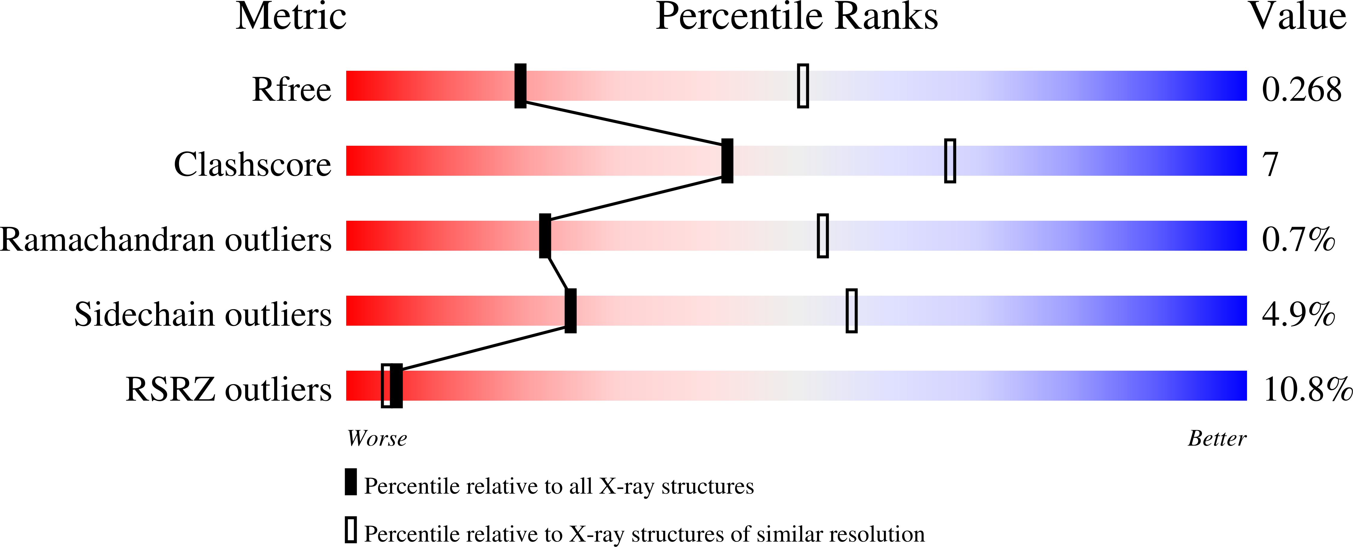
wwPDB Validation   3D Report Full Report


This is version 1.2 of the entry. See complete history


Literature

HU multimerization shift controls nucleoid compaction.

Hammel, M.Amlanjyoti, D.Reyes, F.E.Chen, J.H.Parpana, R.Tang, H.Y.Larabell, C.A.Tainer, J.A.Adhya, S.

(2016) Sci Adv 2: e1600650-e1600650

  • DOI: https://doi.org/10.1126/sciadv.1600650
  • Primary Citation of Related Structures:  
    4YEW, 4YEX, 4YEY, 4YF0, 4YFH, 4YFT

  • PubMed Abstract: 

    Molecular mechanisms controlling functional bacterial chromosome (nucleoid) compaction and organization are surprisingly enigmatic but partly depend on conserved, histone-like proteins HUαα and HUαβ and their interactions that span the nanoscale and mesoscale from protein-DNA complexes to the bacterial chromosome and nucleoid structure. We determined the crystal structures of these chromosome-associated proteins in complex with native duplex DNA. Distinct DNA binding modes of HUαα and HUαβ elucidate fundamental features of bacterial chromosome packing that regulate gene transcription. By combining crystal structures with solution x-ray scattering results, we determined architectures of HU-DNA nucleoproteins in solution under near-physiological conditions. These macromolecular conformations and interactions result in contraction at the cellular level based on in vivo imaging of native unlabeled nucleoid by soft x-ray tomography upon HUβ and ectopic HUα38 expression. Structural characterization of charge-altered HUαα-DNA complexes reveals an HU molecular switch that is suitable for condensing nucleoid and reprogramming noninvasive Escherichia coli into an invasive form. Collective findings suggest that shifts between networking and cooperative and noncooperative DNA-dependent HU multimerization control DNA compaction and supercoiling independently of cellular topoisomerase activity. By integrating x-ray crystal structures, x-ray scattering, mutational tests, and x-ray imaging that span from protein-DNA complexes to the bacterial chromosome and nucleoid structure, we show that defined dynamic HU interaction networks can promote nucleoid reorganization and transcriptional regulation as efficient general microbial mechanisms to help synchronize genetic responses to cell cycle, changing environments, and pathogenesis.


  • Organizational Affiliation

    Molecular Biophysics and Integrated Bioimaging, Lawrence Berkeley National Laboratory, Berkeley, CA 94720, USA.


Macromolecules

Find similar proteins by:  (by identity cutoff)  |  3D Structure
Entity ID: 1
MoleculeChains Sequence LengthOrganismDetailsImage
DNA-binding protein HU-beta90Escherichia coliMutation(s): 0 
Gene Names: hupB
UniProt
Find proteins for P0ACF4 (Escherichia coli (strain K12))
Explore P0ACF4 
Go to UniProtKB:  P0ACF4
Entity Groups  
Sequence Clusters30% Identity50% Identity70% Identity90% Identity95% Identity100% Identity
UniProt GroupP0ACF4
Sequence Annotations
Expand
  • Reference Sequence
Find similar proteins by:  (by identity cutoff)  |  3D Structure
Entity ID: 4
MoleculeChains Sequence LengthOrganismDetailsImage
DNA-binding protein HU-alphaD [auth C]90Escherichia coliMutation(s): 0 
Gene Names: hupA
UniProt
Find proteins for P0ACF0 (Escherichia coli (strain K12))
Explore P0ACF0 
Go to UniProtKB:  P0ACF0
Entity Groups  
Sequence Clusters30% Identity50% Identity70% Identity90% Identity95% Identity100% Identity
UniProt GroupP0ACF0
Sequence Annotations
Expand
  • Reference Sequence

Find similar nucleic acids by:  Sequence   |   3D Structure  

Entity ID: 2
MoleculeChains LengthOrganismImage
synthetic DNA strand9Escherichia coli
Sequence Annotations
Expand
  • Reference Sequence

Find similar nucleic acids by:  Sequence   |   3D Structure  

Entity ID: 3
MoleculeChains LengthOrganismImage
synthetic DNA strandC [auth D]11Escherichia coli
Sequence Annotations
Expand
  • Reference Sequence
Experimental Data & Validation

Experimental Data

  • Method: X-RAY DIFFRACTION
  • Resolution: 2.91 Å
  • R-Value Free: 0.251 
  • R-Value Work: 0.215 
  • R-Value Observed: 0.216 
  • Space Group: P 42 21 2
Unit Cell:
Length ( Å )Angle ( ˚ )
a = 83.036α = 90
b = 83.036β = 90
c = 63.114γ = 90
Software Package:
Software NamePurpose
BUSTER-TNTrefinement
PDB_EXTRACTdata extraction
Aimlessdata scaling
BUSTERrefinement
XDSdata reduction
MOLREPphasing

Structure Validation

View Full Validation Report


Entry History 

Deposition Data

Revision History  (Full details and data files)

  • Version 1.0: 2016-06-29
    Type: Initial release
  • Version 1.1: 2019-02-20
    Changes: Data collection, Database references, Derived calculations
  • Version 1.2: 2023-09-27
    Changes: Data collection, Database references, Refinement description