4YDR

Crystal structure of oxidized homoserine dehydrogenase of Sulfolobus tokodaii


Experimental Data Snapshot

  • Method: X-RAY DIFFRACTION
  • Resolution: 1.60 Å
  • R-Value Free: 0.228 
  • R-Value Work: 0.197 
  • R-Value Observed: 0.198 

wwPDB Validation   3D Report Full Report


This is version 1.1 of the entry. See complete history


Literature

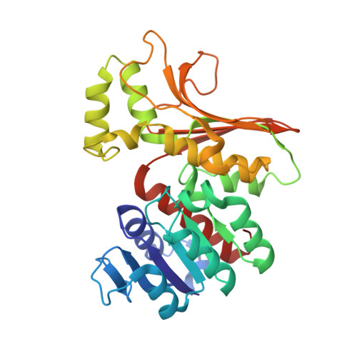
Structural insight into activation of homoserine dehydrogenase from the archaeonSulfolobus tokodaiivia reduction.

Tomonaga, Y.Kaneko, R.Goto, M.Ohshima, T.Yoshimune, K.

(2015) Biochem Biophys Rep 3: 14-17

  • DOI: https://doi.org/10.1016/j.bbrep.2015.07.006
  • Primary Citation of Related Structures:  
    4YDR, 5AVO

  • PubMed Abstract: 

    Homoserine dehydrogenase (HSD; 305 amino acid residues) catalyzes an NAD(P)-dependent reversible reaction between l-homoserine and aspartate 4-semialdehyde and is involved in the aspartate pathway. HSD from the hyperthermophilic archaeon Sulfolobus tokodaii was markedly activated (2.5-fold) by the addition of 0.8 mM dithiothreitol. The crystal structure of the homodimer indicated that the activation was caused by cleavage of the disulfide bond formed between two cysteine residues (C303) in the C-terminal regions of the two subunits.


  • Organizational Affiliation

    Department of Applied Molecular Chemistry, Graduate School of Industrial Technology, Nihon University, 1-2-1, Izumichou, Narashino, Chiba 275-8575, Japan.


Macromolecules
Find similar proteins by:  (by identity cutoff)  |  3D Structure
Entity ID: 1
MoleculeChains Sequence LengthOrganismDetailsImage
Homoserine dehydrogenase
A, B
304Sulfurisphaera tokodaii str. 7Mutation(s): 0 
Gene Names: homSTK_15190
EC: 1.1.1.3
UniProt
Find proteins for F9VNG5 (Sulfurisphaera tokodaii (strain DSM 16993 / JCM 10545 / NBRC 100140 / 7))
Explore F9VNG5 
Go to UniProtKB:  F9VNG5
Entity Groups  
Sequence Clusters30% Identity50% Identity70% Identity90% Identity95% Identity100% Identity
UniProt GroupF9VNG5
Sequence Annotations
Expand
  • Reference Sequence
Small Molecules
Ligands 1 Unique
IDChains Name / Formula / InChI Key2D Diagram3D Interactions
NA
Query on NA

Download Ideal Coordinates CCD File 
C [auth A]SODIUM ION
Na
FKNQFGJONOIPTF-UHFFFAOYSA-N
Experimental Data & Validation

Experimental Data

  • Method: X-RAY DIFFRACTION
  • Resolution: 1.60 Å
  • R-Value Free: 0.228 
  • R-Value Work: 0.197 
  • R-Value Observed: 0.198 
  • Space Group: P 1 21 1
Unit Cell:
Length ( Å )Angle ( ˚ )
a = 57.818α = 90
b = 78.909β = 105.79
c = 65.814γ = 90
Software Package:
Software NamePurpose
REFMACrefinement
HKL-2000data reduction
HKL-2000data scaling
PHASERphasing

Structure Validation

View Full Validation Report


Entry History 

Deposition Data

Revision History  (Full details and data files)

  • Version 1.0: 2015-11-11
    Type: Initial release
  • Version 1.1: 2019-12-18
    Changes: Data collection, Database references, Derived calculations