4Y5R

Crystal Structure of a T67A MauG/pre-Methylamine Dehydrogenase Complex


Experimental Data Snapshot

  • Method: X-RAY DIFFRACTION
  • Resolution: 2.80 Å
  • R-Value Free: 0.256 
  • R-Value Work: 0.193 
  • R-Value Observed: 0.196 

wwPDB Validation   3D Report Full Report


Ligand Structure Quality Assessment 


This is version 1.6 of the entry. See complete history


Literature

A T67A mutation in the proximal pocket of the high-spin heme of MauG stabilizes formation of a mixed-valent Fe(II)/Fe(III) state and enhances charge resonance stabilization of the bis-Fe(IV) state.

Shin, S.Feng, M.Li, C.Williamson, H.R.Choi, M.Wilmot, C.M.Davidson, V.L.

(2015) Biochim Biophys Acta 1847: 709-716

  • DOI: https://doi.org/10.1016/j.bbabio.2015.04.008
  • Primary Citation of Related Structures:  
    4Y5R

  • PubMed Abstract: 

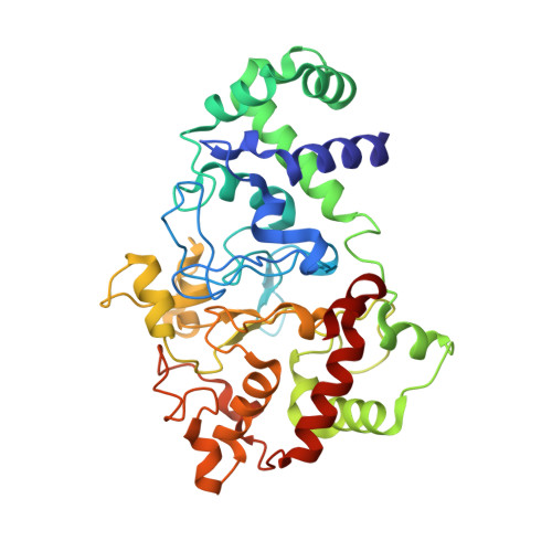
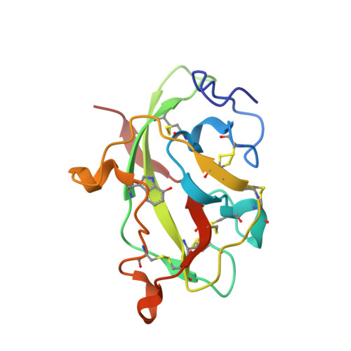
    The diheme enzyme MauG catalyzes a six-electron oxidation required for posttranslational modification of a precursor of methylamine dehydrogenase (preMADH) to complete the biosynthesis of its protein-derived tryptophan tryptophylquinone (TTQ) cofactor. One heme is low-spin with ligands provided by His205 and Tyr294, and the other is high-spin with a ligand provided by His35. The side chain methyl groups of Thr67 and Leu70 are positioned at a distance of 3.4Å on either side of His35, maintaining a hydrophobic environment in the proximal pocket of the high-spin heme and restricting the movement of this ligand. Mutation of Thr67 to Ala in the proximal pocket of the high-spin heme prevented reduction of the low-spin heme by dithionite, yielding a mixed-valent state. The mutation also enhanced the stabilization of the charge-resonance-transition of the high-valent bis-FeIV state that is generated by addition of H2O2. The rates of electron transfer from TTQ biosynthetic intermediates to the high-valent form of T67A MauG were similar to that of wild-type MauG. These results are compared to those previously reported for mutation of residues in the distal pocket of the high-spin heme that also affected the redox properties and charge resonance transition stabilization of the high-valent state of the hemes. However, given the position of residue 67, the structure of the variant protein and the physical nature of the T67A mutation, the basis for the effects of the T67A mutation must be different from those of the mutations of the residues in the distal heme pocket.


  • Organizational Affiliation

    Department of Bioengineering and Biotechnology, College of Engineering, Chonnam National University, Chonnam, South Korea.


Macromolecules
Find similar proteins by:  (by identity cutoff)  |  3D Structure
Entity ID: 1
MoleculeChains Sequence LengthOrganismDetailsImage
Methylamine utilization protein MauG
A, B
355Paracoccus denitrificans PD1222Mutation(s): 1 
Gene Names: mauGPden_4736
EC: 1
UniProt
Find proteins for Q51658 (Paracoccus denitrificans (strain Pd 1222))
Explore Q51658 
Go to UniProtKB:  Q51658
Entity Groups  
Sequence Clusters30% Identity50% Identity70% Identity90% Identity95% Identity100% Identity
UniProt GroupQ51658
Sequence Annotations
Expand
  • Reference Sequence
Find similar proteins by:  (by identity cutoff)  |  3D Structure
Entity ID: 2
MoleculeChains Sequence LengthOrganismDetailsImage
Methylamine dehydrogenase light chain
C, E
125Paracoccus denitrificansMutation(s): 0 
Gene Names: mauA
EC: 1.4.9.1
UniProt
Find proteins for P22619 (Paracoccus denitrificans)
Explore P22619 
Go to UniProtKB:  P22619
Entity Groups  
Sequence Clusters30% Identity50% Identity70% Identity90% Identity95% Identity100% Identity
UniProt GroupP22619
Sequence Annotations
Expand
  • Reference Sequence
Find similar proteins by:  (by identity cutoff)  |  3D Structure
Entity ID: 3
MoleculeChains Sequence LengthOrganismDetailsImage
Methylamine dehydrogenase heavy chain
D, F
376Paracoccus denitrificans PD1222Mutation(s): 0 
Gene Names: Pden_4730
EC: 1.4.9.1
UniProt
Find proteins for A1BB97 (Paracoccus denitrificans (strain Pd 1222))
Explore A1BB97 
Go to UniProtKB:  A1BB97
Entity Groups  
Sequence Clusters30% Identity50% Identity70% Identity90% Identity95% Identity100% Identity
UniProt GroupA1BB97
Sequence Annotations
Expand
  • Reference Sequence
Experimental Data & Validation

Experimental Data

  • Method: X-RAY DIFFRACTION
  • Resolution: 2.80 Å
  • R-Value Free: 0.256 
  • R-Value Work: 0.193 
  • R-Value Observed: 0.196 
  • Space Group: P 1
Unit Cell:
Length ( Å )Angle ( ˚ )
a = 55.53α = 109.94
b = 83.52β = 91.54
c = 107.78γ = 105.78
Software Package:
Software NamePurpose
REFMACrefinement
Aimlessdata scaling
PDB_EXTRACTdata extraction
XDSdata scaling
REFMACphasing

Structure Validation

View Full Validation Report


Ligand Structure Quality Assessment 


Entry History & Funding Information

Deposition Data


Funding OrganizationLocationGrant Number
National Institutes of Health/National Institute of General Medical Sciences (NIH/NIGMS)United States--

Revision History  (Full details and data files)

  • Version 1.0: 2015-06-17
    Type: Initial release
  • Version 1.1: 2016-07-20
    Changes: Data collection
  • Version 1.2: 2017-09-20
    Changes: Author supporting evidence, Derived calculations, Refinement description
  • Version 1.3: 2019-07-17
    Changes: Data collection, Derived calculations
  • Version 1.4: 2019-12-25
    Changes: Author supporting evidence
  • Version 1.5: 2023-09-27
    Changes: Data collection, Database references, Derived calculations, Refinement description
  • Version 1.6: 2023-11-15
    Changes: Data collection