4XPL

The crystal structure of Campylobacter jejuni N-acetyltransferase PseH in complex with acetyl coenzyme A


Experimental Data Snapshot

  • Method: X-RAY DIFFRACTION
  • Resolution: 1.95 Å
  • R-Value Free: 0.229 
  • R-Value Work: 0.188 
  • R-Value Observed: 0.190 

wwPDB Validation   3D Report Full Report


Ligand Structure Quality Assessment 


This is version 1.2 of the entry. See complete history


Literature

Structural analysis of PseH, the Campylobacter jejuni N-acetyltransferase involved in bacterial O-linked glycosylation.

Song, W.S.Nam, M.S.Namgung, B.Yoon, S.I.

(2015) Biochem Biophys Res Commun 458: 843-848

  • DOI: https://doi.org/10.1016/j.bbrc.2015.02.041
  • Primary Citation of Related Structures:  
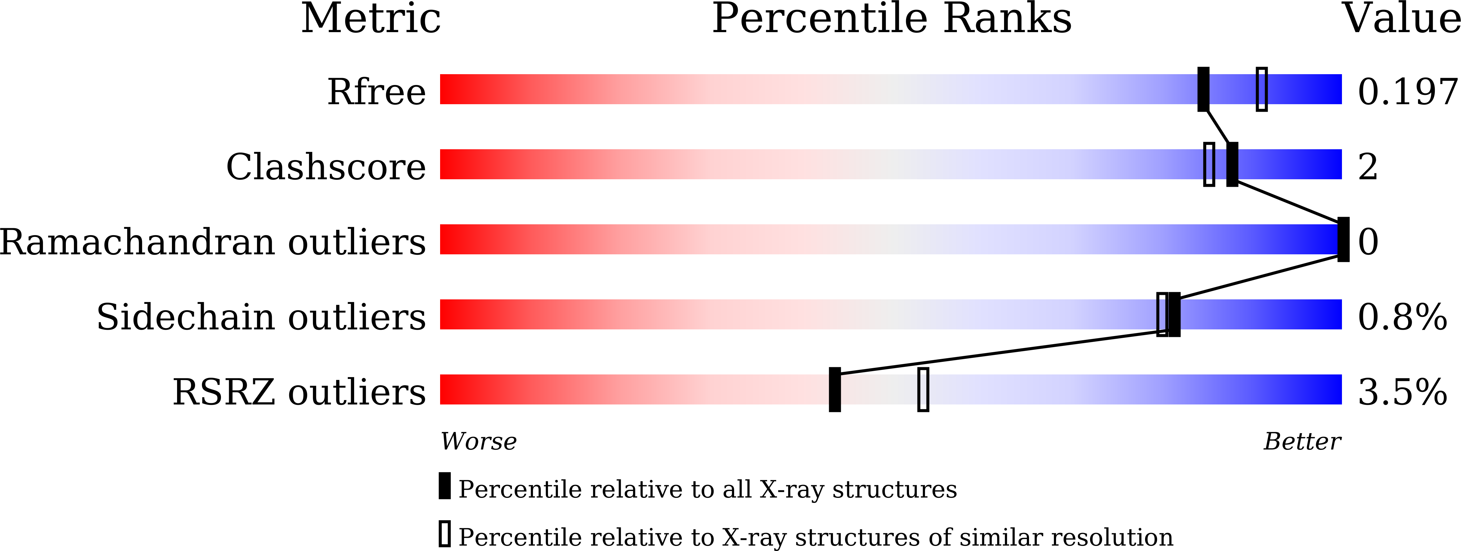
    4XPK, 4XPL

  • PubMed Abstract: 

    Campylobacter jejuni is a bacterium that uses flagella for motility and causes worldwide acute gastroenteritis in humans. The C. jejuni N-acetyltransferase PseH (cjPseH) is responsible for the third step in flagellin O-linked glycosylation and plays a key role in flagellar formation and motility. cjPseH transfers an acetyl group from an acetyl donor, acetyl coenzyme A (AcCoA), to the amino group of UDP-4-amino-4,6-dideoxy-N-acetyl-β-L-altrosamine to produce UDP-2,4-diacetamido-2,4,6-trideoxy-β-L-altropyranose. To elucidate the catalytic mechanism of cjPseH, crystal structures of cjPseH alone and in complex with AcCoA were determined at 1.95 Å resolution. cjPseH folds into a single-domain structure of a central β-sheet decorated by four α-helices with two continuously connected grooves. A deep groove (groove-A) accommodates the AcCoA molecule. Interestingly, the acetyl end of AcCoA points toward an open space in a neighboring shallow groove (groove-S), which is occupied by extra electron density that potentially serves as a pseudosubstrate, suggesting that the groove-S may provide a substrate-binding site. Structure-based comparative analysis suggests that cjPseH utilizes a unique catalytic mechanism of acetylation that has not been observed in other glycosylation-associated acetyltransferases. Thus, our studies on cjPseH will provide valuable information for the design of new antibiotics to treat C. jejuni-induced gastroenteritis.


  • Organizational Affiliation

    Department of Systems Immunology, College of Biomedical Science, Kangwon National University, Chuncheon 200-701, Republic of Korea.


Macromolecules
Find similar proteins by:  (by identity cutoff)  |  3D Structure
Entity ID: 1
MoleculeChains Sequence LengthOrganismDetailsImage
N-Acetyltransferase, PseH163Campylobacter jejuni subsp. jejuni PT14Mutation(s): 8 
Gene Names: A911_06385
UniProt
Find proteins for A0A0J9X276 (Campylobacter jejuni subsp. jejuni PT14)
Explore A0A0J9X276 
Go to UniProtKB:  A0A0J9X276
Entity Groups  
Sequence Clusters30% Identity50% Identity70% Identity90% Identity95% Identity100% Identity
UniProt GroupA0A0J9X276
Sequence Annotations
Expand
  • Reference Sequence
Small Molecules
Ligands 1 Unique
IDChains Name / Formula / InChI Key2D Diagram3D Interactions
ACO
Query on ACO

Download Ideal Coordinates CCD File 
B [auth A]ACETYL COENZYME *A
C23 H38 N7 O17 P3 S
ZSLZBFCDCINBPY-ZSJPKINUSA-N
Experimental Data & Validation

Experimental Data

  • Method: X-RAY DIFFRACTION
  • Resolution: 1.95 Å
  • R-Value Free: 0.229 
  • R-Value Work: 0.188 
  • R-Value Observed: 0.190 
  • Space Group: P 21 21 2
Unit Cell:
Length ( Å )Angle ( ˚ )
a = 42.421α = 90
b = 110.495β = 90
c = 40.785γ = 90
Software Package:
Software NamePurpose
HKL-2000data reduction
REFMACrefinement
PDB_EXTRACTdata extraction
HKL-2000data scaling
PHASERphasing

Structure Validation

View Full Validation Report


Ligand Structure Quality Assessment 


Entry History 

Deposition Data

Revision History  (Full details and data files)

  • Version 1.0: 2015-03-18
    Type: Initial release
  • Version 1.1: 2015-03-25
    Changes: Database references
  • Version 1.2: 2023-11-08
    Changes: Data collection, Database references, Derived calculations, Refinement description, Source and taxonomy