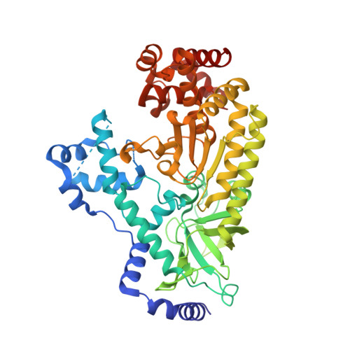
4XM6

Anthrax toxin lethal factor with ligand-induced binding pocket


Experimental Data Snapshot

  • Method: X-RAY DIFFRACTION
  • Resolution: 2.35 Å
  • R-Value Free: 0.246 
  • R-Value Work: 0.205 
  • R-Value Observed: 0.207 

wwPDB Validation   3D Report Full Report


Ligand Structure Quality Assessment 


This is version 1.5 of the entry. See complete history


Literature

Ligand-induced expansion of the S1' site in the anthrax toxin lethal factor.

Maize, K.M.Kurbanov, E.K.Johnson, R.L.Amin, E.A.Finzel, B.C.

(2015) FEBS Lett 589: 3836-3841

  • DOI: https://doi.org/10.1016/j.febslet.2015.11.005
  • Primary Citation of Related Structures:  
    4XM6, 4XM7, 4XM8

  • PubMed Abstract: 

    The Bacillus anthracis lethal factor (LF) is one component of a tripartite exotoxin partly responsible for persistent anthrax cytotoxicity after initial bacterial infection. Inhibitors of the zinc metalloproteinase have been investigated as potential therapeutic agents, but LF is a challenging target because inhibitors lack sufficient selectivity or possess poor pharmaceutical properties. These structural studies reveal an alternate conformation of the enzyme, induced upon binding of specific inhibitors, that opens a previously unobserved deep pocket termed S1'(∗) which might afford new opportunities to design selective inhibitors that target this subsite.


  • Organizational Affiliation

    Department of Medicinal Chemistry, University of Minnesota, 308 Harvard St SE, 8-101 Weaver-Densford Hall, Minneapolis, MN 55455, United States.


Macromolecules
Find similar proteins by:  (by identity cutoff)  |  3D Structure
Entity ID: 1
MoleculeChains Sequence LengthOrganismDetailsImage
Lethal factor519Bacillus anthracisMutation(s): 1 
Gene Names: lefpXO1-107BXA0172GBAA_pXO1_0172
EC: 3.4.24.83
UniProt
Find proteins for P15917 (Bacillus anthracis)
Explore P15917 
Go to UniProtKB:  P15917
Entity Groups  
Sequence Clusters30% Identity50% Identity70% Identity90% Identity95% Identity100% Identity
UniProt GroupP15917
Sequence Annotations
Expand
  • Reference Sequence
Small Molecules
Binding Affinity Annotations 
IDSourceBinding Affinity
41R Binding MOAD:  4XM6 IC50: 9.00e+4 (nM) from 1 assay(s)
Experimental Data & Validation

Experimental Data

  • Method: X-RAY DIFFRACTION
  • Resolution: 2.35 Å
  • R-Value Free: 0.246 
  • R-Value Work: 0.205 
  • R-Value Observed: 0.207 
  • Space Group: P 21 21 21
Unit Cell:
Length ( Å )Angle ( ˚ )
a = 57.936α = 90
b = 76.922β = 90
c = 138.576γ = 90
Software Package:
Software NamePurpose
HKL-2000data scaling
HKL-2000data reduction
PHASERphasing
PHENIXrefinement
PDB_EXTRACTdata extraction

Structure Validation

View Full Validation Report


Ligand Structure Quality Assessment 


Entry History & Funding Information

Deposition Data


Funding OrganizationLocationGrant Number
National Institutes of Health/National Institute Of Allergy and Infectious Diseases (NIH/NIAID)United StatesAI091790-01
Minnesota Dept. of Employment and Economic DevelopmentUnited StatesSPAP-06-0014-P-FY07

Revision History  (Full details and data files)

  • Version 1.0: 2015-11-11
    Type: Initial release
  • Version 1.1: 2015-12-02
    Changes: Database references
  • Version 1.2: 2015-12-30
    Changes: Database references
  • Version 1.3: 2017-09-27
    Changes: Author supporting evidence, Database references, Derived calculations
  • Version 1.4: 2019-12-11
    Changes: Author supporting evidence
  • Version 1.5: 2023-09-27
    Changes: Data collection, Database references, Refinement description