


`The Hunt for Serendipitous Allosteric Sites: Discovery of a novel allosteric inhibitor of the bacterial sialidase NanB
Rogers, G.W., Brear, P., Yang, L., Chen, A.S., Mitchell, J.B.O., Taylor, G.L., Westwood, N.J.To be published.
Experimental Data Snapshot
Entity ID: 1 | |||||
|---|---|---|---|---|---|
| Molecule | Chains | Sequence Length | Organism | Details | Image |
| Sialidase B | 658 | Streptococcus pneumoniae TIGR4 | Mutation(s): 1 Gene Names: nanB, SP_1687 EC: 3.2.1.18 | 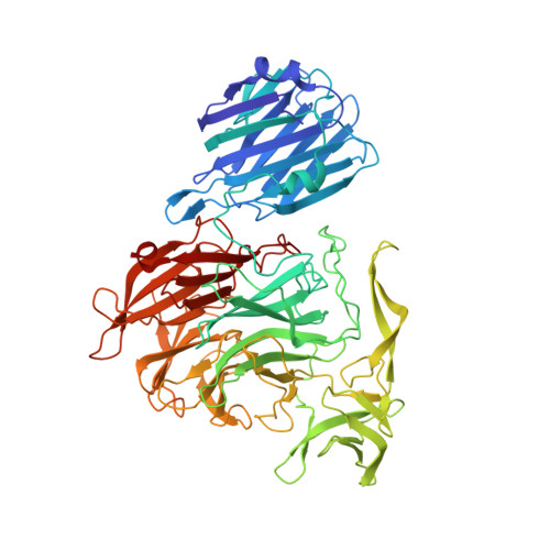 | |
UniProt | |||||
Find proteins for Q54727 (Streptococcus pneumoniae serotype 4 (strain ATCC BAA-334 / TIGR4)) Explore Q54727 Go to UniProtKB: Q54727 | |||||
Entity Groups | |||||
| Sequence Clusters | 30% Identity50% Identity70% Identity90% Identity95% Identity100% Identity | ||||
| UniProt Group | Q54727 | ||||
Sequence AnnotationsExpand | |||||
| |||||
| Ligands 4 Unique | |||||
|---|---|---|---|---|---|
| ID | Chains | Name / Formula / InChI Key | 2D Diagram | 3D Interactions | |
| SFJ Query on SFJ | F [auth A] | (2R,3R,4R,5R,6R)-5-acetamido-2,3-difluoro-4-hydroxy-6-[(1R,2R)-1,2,3-trihydroxypropyl]tetrahydro-2H-pyran-2-carboxylic
acid C11 H17 F2 N O8 HMALKZAXODOYOP-DAXAGCIGSA-N |  | ||
| OPX Query on OPX | B [auth A] | (1s,3R,4S)-1-[(cyclohexylamino)methyl]-3,4-dihydroxycyclopentanesulfonic acid C12 H23 N O5 S RMLVAYWMDDDXFB-ZSBIGDGJSA-N |  | ||
| PO4 Query on PO4 | C [auth A] | PHOSPHATE ION O4 P NBIIXXVUZAFLBC-UHFFFAOYSA-K |  | ||
| DMS Query on DMS | D [auth A], E [auth A] | DIMETHYL SULFOXIDE C2 H6 O S IAZDPXIOMUYVGZ-UHFFFAOYSA-N |  | ||
| Length ( Å ) | Angle ( ˚ ) |
|---|---|
| a = 76.586 | α = 90 |
| b = 82.556 | β = 90 |
| c = 116.654 | γ = 90 |
| Software Name | Purpose |
|---|---|
| PHENIX | refinement |
| HKL-2000 | data reduction |
| PHASER | phasing |
| PDB_EXTRACT | data extraction |
| HKL-2000 | data scaling |
| Funding Organization | Location | Grant Number |
|---|---|---|
| Biotechnology and Biological Sciences Research Council | United Kingdom | -- |
| SULSA | United Kingdom | -- |
RCSB PDB (citation) is hosted by
RCSB PDB is a member of the

