4XGO

Crystal structure of leucine-rich repeat domain of APL1B


Experimental Data Snapshot

  • Method: X-RAY DIFFRACTION
  • Resolution: 1.74 Å
  • R-Value Free: 0.220 
  • R-Value Work: 0.187 
  • R-Value Observed: 0.188 

wwPDB Validation   3D Report Full Report


This is version 2.0 of the entry. See complete history


Literature

Biophysical Analysis of Anopheles gambiae Leucine-Rich Repeat Proteins APL1A1, APLB and APL1C and Their Interaction with LRIM1.

Williams, M.Summers, B.J.Baxter, R.H.

(2015) PLoS One 10: e0118911-e0118911

  • DOI: https://doi.org/10.1371/journal.pone.0118911
  • Primary Citation of Related Structures:  
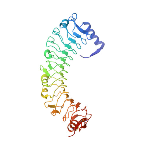
    4XGO

  • PubMed Abstract: 

    Natural infection of Anopheles gambiae by malaria-causing Plasmodium parasites is significantly influenced by the APL1 genetic locus. The locus contains three closely related leucine-rich repeat (LRR) genes, APL1A, APL1B and APL1C. Multiple studies have reported the participation of APL1A-C in the immune response of A. gambiae to invasion by both rodent and human Plasmodium isolates. APL1C forms a heterodimer with the related LRR protein LRIM1 via a C-terminal coiled-coil domain that is also present in APL1A and APL1B. The LRIM1/APL1C heterodimer protects A. gambiae from infection by binding the complement-like protein TEP1 to form a stable and active immune complex. Here we report solution x-ray scatting data for the LRIM1/APL1C heterodimer, the oligomeric state of LRIM1/APL1 LRR domains in solution and the crystal structure of the APL1B LRR domain. The LRIM1/APL1C heterodimeric complex has a flexible and extended structure in solution. In contrast to the APL1A, APL1C and LRIM1 LRR domains, the APL1B LRR domain is a homodimer. The crystal structure of APL1B-LRR shows that the homodimer is formed by an N-terminal helix that complements for the absence of an N-terminal capping motif in APL1B, which is a unique distinction within the LRIM1/APL1 protein family. Full-length APL1A1 and APL1B form a stable complex with LRIM1. These results support a model in which APL1A1, APL1B and APL1C can all form an extended, flexible heterodimer with LRIM1, providing a repertoire of functional innate immune complexes to protect A. gambiae from a diverse array of pathogens.


  • Organizational Affiliation

    Department of Chemistry and Molecular Biophysics & Biochemistry, Yale University, New Haven, Connecticut, United States of America.


Macromolecules
Find similar proteins by:  (by identity cutoff)  |  3D Structure
Entity ID: 1
MoleculeChains Sequence LengthOrganismDetailsImage
Anopheles Plasmodium-responsive Leucine-rich repeat protein 1B
A, B
356Anopheles gambiaeMutation(s): 0 
Gene Names: APL1B
UniProt
Find proteins for F2YBL9 (Anopheles gambiae)
Explore F2YBL9 
Go to UniProtKB:  F2YBL9
Entity Groups  
Sequence Clusters30% Identity50% Identity70% Identity90% Identity95% Identity100% Identity
UniProt GroupF2YBL9
Sequence Annotations
Expand
  • Reference Sequence
Oligosaccharides

Help

Entity ID: 2
MoleculeChains Length2D Diagram Glycosylation3D Interactions
2-acetamido-2-deoxy-beta-D-glucopyranose-(1-4)-2-acetamido-2-deoxy-beta-D-glucopyranose
C, D
2N-Glycosylation
Glycosylation Resources
GlyTouCan:  G42666HT
GlyCosmos:  G42666HT
GlyGen:  G42666HT
Small Molecules
Ligands 5 Unique
IDChains Name / Formula / InChI Key2D Diagram3D Interactions
PO4
Query on PO4

Download Ideal Coordinates CCD File 
E [auth A],
O [auth B],
P [auth B]
PHOSPHATE ION
O4 P
NBIIXXVUZAFLBC-UHFFFAOYSA-K
GOL
Query on GOL

Download Ideal Coordinates CCD File 
G [auth A]
H [auth A]
I [auth A]
J [auth A]
K [auth A]
G [auth A],
H [auth A],
I [auth A],
J [auth A],
K [auth A],
L [auth A]
GLYCEROL
C3 H8 O3
PEDCQBHIVMGVHV-UHFFFAOYSA-N
ACT
Query on ACT

Download Ideal Coordinates CCD File 
F [auth A],
N [auth B]
ACETATE ION
C2 H3 O2
QTBSBXVTEAMEQO-UHFFFAOYSA-M
CL
Query on CL

Download Ideal Coordinates CCD File 
M [auth A],
Q [auth B]
CHLORIDE ION
Cl
VEXZGXHMUGYJMC-UHFFFAOYSA-M
NA
Query on NA

Download Ideal Coordinates CCD File 
R [auth B]SODIUM ION
Na
FKNQFGJONOIPTF-UHFFFAOYSA-N
Experimental Data & Validation

Experimental Data

  • Method: X-RAY DIFFRACTION
  • Resolution: 1.74 Å
  • R-Value Free: 0.220 
  • R-Value Work: 0.187 
  • R-Value Observed: 0.188 
  • Space Group: P 21 21 21
Unit Cell:
Length ( Å )Angle ( ˚ )
a = 64.423α = 90
b = 74.956β = 90
c = 214.37γ = 90
Software Package:
Software NamePurpose
REFMACrefinement
HKL-2000data reduction
HKL-2000data scaling
PHASERphasing

Structure Validation

View Full Validation Report


Entry History 

Deposition Data

Revision History  (Full details and data files)

  • Version 1.0: 2015-04-01
    Type: Initial release
  • Version 2.0: 2020-07-29
    Type: Remediation
    Reason: Carbohydrate remediation
    Changes: Atomic model, Data collection, Derived calculations, Source and taxonomy, Structure summary