4XB2

Hyperthermophilic archaeal homoserine dehydrogenase mutant in complex with NADPH


Experimental Data Snapshot

  • Method: X-RAY DIFFRACTION
  • Resolution: 2.43 Å
  • R-Value Free: 0.223 
  • R-Value Work: 0.164 
  • R-Value Observed: 0.167 

wwPDB Validation   3D Report Full Report


Ligand Structure Quality Assessment 


This is version 2.0 of the entry. See complete history


Literature

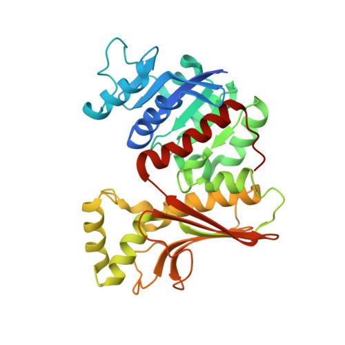
Crystal Structures of a Hyperthermophilic Archaeal Homoserine Dehydrogenase Suggest a Novel Cofactor Binding Mode for Oxidoreductases.

Hayashi, J.Inoue, S.Kim, K.Yoneda, K.Kawarabayasi, Y.Ohshima, T.Sakuraba, H.

(2015) Sci Rep 5: 11674-11674

  • DOI: https://doi.org/10.1038/srep11674
  • Primary Citation of Related Structures:  
    4XB1, 4XB2

  • PubMed Abstract: 

    NAD(P)-dependent dehydrogenases differ according to their coenzyme preference: some prefer NAD, others NADP, and still others exhibit dual cofactor specificity. The structure of a newly identified archaeal homoserine dehydrogenase showed this enzyme to have a strong preference for NADP. However, NADP did not act as a cofactor with this enzyme, but as a strong inhibitor of NAD-dependent homoserine oxidation. Structural analysis and site-directed mutagenesis showed that the large number of interactions between the cofactor and the enzyme are responsible for the lack of reactivity of the enzyme towards NADP. This observation suggests this enzyme exhibits a new variation on cofactor binding to a dehydrogenase: very strong NADP binding that acts as an obstacle to NAD(P)-dependent dehydrogenase catalytic activity.


  • Organizational Affiliation

    Department of Applied Biological Science, Faculty of Agriculture, Kagawa University, Ikenobe 2393, Miki-cho, Kagawa 761-0795, Japan.


Macromolecules
Find similar proteins by:  (by identity cutoff)  |  3D Structure
Entity ID: 1
MoleculeChains Sequence LengthOrganismDetailsImage
319aa long hypothetical homoserine dehydrogenase
A, B
335Pyrococcus horikoshii OT3Mutation(s): 1 
Gene Names: PH1075
UniProt
Find proteins for O58802 (Pyrococcus horikoshii (strain ATCC 700860 / DSM 12428 / JCM 9974 / NBRC 100139 / OT-3))
Explore O58802 
Go to UniProtKB:  O58802
Entity Groups  
Sequence Clusters30% Identity50% Identity70% Identity90% Identity95% Identity100% Identity
UniProt GroupO58802
Sequence Annotations
Expand
  • Reference Sequence
Experimental Data & Validation

Experimental Data

  • Method: X-RAY DIFFRACTION
  • Resolution: 2.43 Å
  • R-Value Free: 0.223 
  • R-Value Work: 0.164 
  • R-Value Observed: 0.167 
  • Space Group: P 1 21 1
Unit Cell:
Length ( Å )Angle ( ˚ )
a = 78.389α = 90
b = 47.811β = 95.54
c = 90.534γ = 90
Software Package:
Software NamePurpose
HKL-2000data reduction
MOLREPphasing
DMrefinement
REFMACrefinement
PDB_EXTRACTdata extraction
HKL-2000data scaling

Structure Validation

View Full Validation Report


Ligand Structure Quality Assessment 


Entry History 

Deposition Data

Revision History  (Full details and data files)

  • Version 1.0: 2015-07-15
    Type: Initial release
  • Version 1.1: 2019-12-25
    Changes: Database references, Derived calculations, Source and taxonomy
  • Version 1.2: 2023-11-08
    Changes: Data collection, Database references, Refinement description
  • Version 2.0: 2023-11-15
    Changes: Atomic model, Data collection