4WXY

PLPS (inactive glutaminase mutant) co-crystallized with glutamine and R5P.


Experimental Data Snapshot

  • Method: X-RAY DIFFRACTION
  • Resolution: 2.70 Å
  • R-Value Free: 0.257 
  • R-Value Work: 0.209 
  • R-Value Observed: 0.211 

wwPDB Validation   3D Report Full Report


This is version 1.3 of the entry. See complete history


Literature

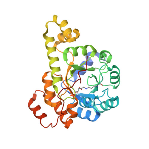
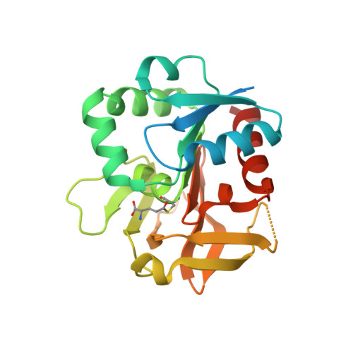
Crystal structures capture three states in the catalytic cycle of a pyridoxal phosphate (PLP) synthase.

Smith, A.M.Brown, W.C.Harms, E.Smith, J.L.

(2015) J Biol Chem 290: 5226-5239

  • DOI: https://doi.org/10.1074/jbc.M114.626382
  • Primary Citation of Related Structures:  
    4WXY, 4WXZ, 4WY0

  • PubMed Abstract: 

    PLP synthase (PLPS) is a remarkable single-enzyme biosynthetic pathway that produces pyridoxal 5'-phosphate (PLP) from glutamine, ribose 5-phosphate, and glyceraldehyde 3-phosphate. The intact enzyme includes 12 synthase and 12 glutaminase subunits. PLP synthesis occurs in the synthase active site by a complicated mechanism involving at least two covalent intermediates at a catalytic lysine. The first intermediate forms with ribose 5-phosphate. The glutaminase subunit is a glutamine amidotransferase that hydrolyzes glutamine and channels ammonia to the synthase active site. Ammonia attack on the first covalent intermediate forms the second intermediate. Glyceraldehyde 3-phosphate reacts with the second intermediate to form PLP. To investigate the mechanism of the synthase subunit, crystal structures were obtained for three intermediate states of the Geobacillus stearothermophilus intact PLPS or its synthase subunit. The structures capture the synthase active site at three distinct steps in its complicated catalytic cycle, provide insights into the elusive mechanism, and illustrate the coordinated motions within the synthase subunit that separate the catalytic states. In the intact PLPS with a Michaelis-like intermediate in the glutaminase active site, the first covalent intermediate of the synthase is fully sequestered within the enzyme by the ordering of a generally disordered 20-residue C-terminal tail. Following addition of ammonia, the synthase active site opens and admits the Lys-149 side chain, which participates in formation of the second intermediate and PLP. Roles are identified for conserved Asp-24 in the formation of the first intermediate and for conserved Arg-147 in the conversion of the first to the second intermediate.


  • Organizational Affiliation

    From the Department of Biological Chemistry, Life Sciences Institute.


Macromolecules
Find similar proteins by:  (by identity cutoff)  |  3D Structure
Entity ID: 1
MoleculeChains Sequence LengthOrganismDetailsImage
Pyridoxal biosynthesis lyase PdxS304Geobacillus kaustophilus HTA426Mutation(s): 0 
Gene Names: pdxSGK0011
EC: 4
UniProt
Find proteins for Q5L3Y2 (Geobacillus kaustophilus (strain HTA426))
Explore Q5L3Y2 
Go to UniProtKB:  Q5L3Y2
Entity Groups  
Sequence Clusters30% Identity50% Identity70% Identity90% Identity95% Identity100% Identity
UniProt GroupQ5L3Y2
Sequence Annotations
Expand
  • Reference Sequence
Find similar proteins by:  (by identity cutoff)  |  3D Structure
Entity ID: 2
MoleculeChains Sequence LengthOrganismDetailsImage
Glutamine amidotransferase subunit PdxT228Geobacillus kaustophilus HTA426Mutation(s): 1 
Gene Names: pdxTGK0012
EC: 2.6
UniProt
Find proteins for Q5L3Y1 (Geobacillus kaustophilus (strain HTA426))
Explore Q5L3Y1 
Go to UniProtKB:  Q5L3Y1
Entity Groups  
Sequence Clusters30% Identity50% Identity70% Identity90% Identity95% Identity100% Identity
UniProt GroupQ5L3Y1
Sequence Annotations
Expand
  • Reference Sequence
Small Molecules
Modified Residues  2 Unique
IDChains TypeFormula2D DiagramParent
L5P
Query on L5P
A
B [auth C]
C [auth E]
D [auth G]
E [auth I]
A,
B [auth C],
C [auth E],
D [auth G],
E [auth I],
F [auth K]
L-PEPTIDE LINKINGC11 H23 N2 O9 PLYS
CYG
Query on CYG
G [auth B]
H [auth D]
I [auth F]
J [auth H]
K [auth J]
G [auth B],
H [auth D],
I [auth F],
J [auth H],
K [auth J],
L
L-PEPTIDE LINKINGC8 H14 N2 O5 SCYS
Experimental Data & Validation

Experimental Data

  • Method: X-RAY DIFFRACTION
  • Resolution: 2.70 Å
  • R-Value Free: 0.257 
  • R-Value Work: 0.209 
  • R-Value Observed: 0.211 
  • Space Group: C 2 2 21
Unit Cell:
Length ( Å )Angle ( ˚ )
a = 141.023α = 90
b = 249.138β = 90
c = 179.656γ = 90
Software Package:
Software NamePurpose
REFMACrefinement
HKL-2000data reduction
PDB_EXTRACTdata extraction
HKL-2000data scaling
BALBESphasing

Structure Validation

View Full Validation Report


Entry History 

Deposition Data

Revision History  (Full details and data files)

  • Version 1.0: 2015-01-14
    Type: Initial release
  • Version 1.1: 2015-01-21
    Changes: Database references
  • Version 1.2: 2015-03-11
    Changes: Database references
  • Version 1.3: 2019-11-27
    Changes: Database references, Derived calculations, Source and taxonomy, Structure summary