4WKM

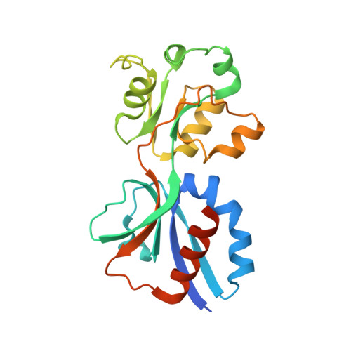
AmpR effector binding domain from Citrobacter freundii bound to UDP-MurNAc-pentapeptide


Experimental Data Snapshot

  • Method: X-RAY DIFFRACTION
  • Resolution: 2.15 Å
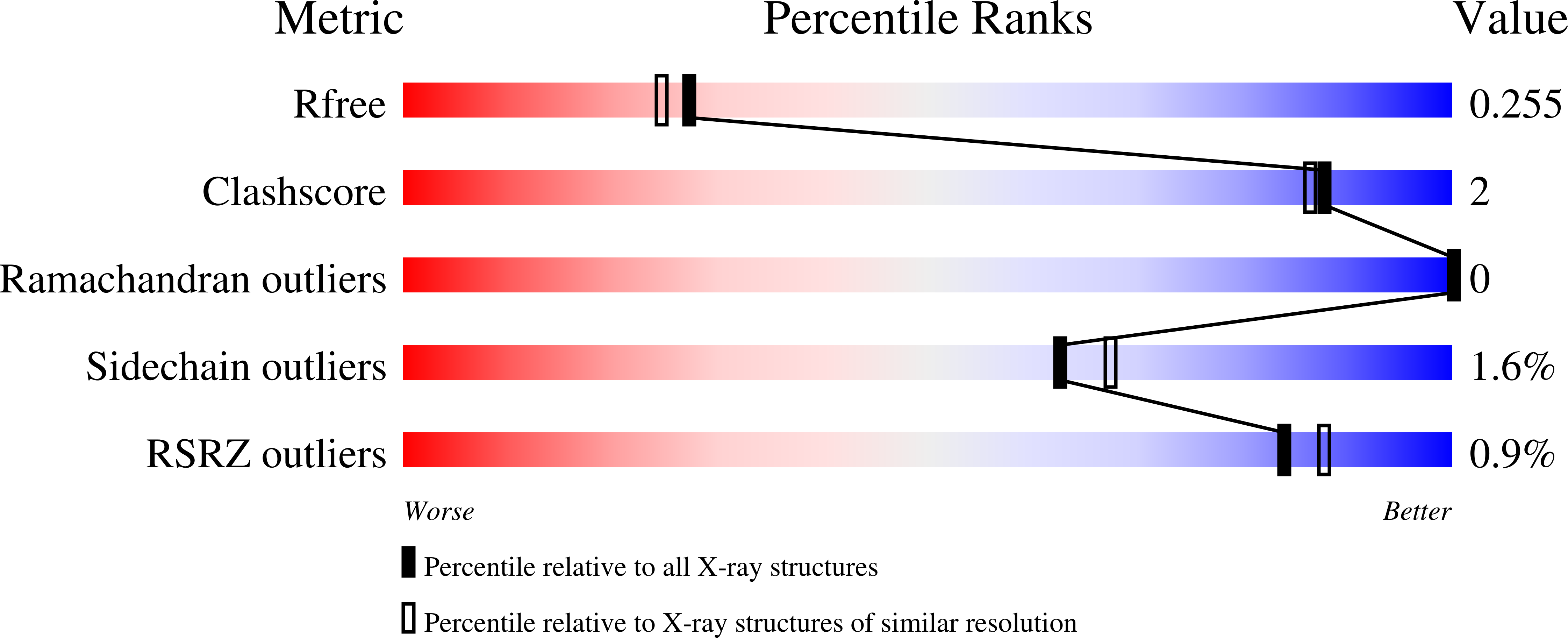
  • R-Value Free: 0.254 
  • R-Value Work: 0.201 
  • R-Value Observed: 0.202 

wwPDB Validation   3D Report Full Report


Ligand Structure Quality Assessment 


This is version 3.0 of the entry. See complete history


Literature

The beta-Lactamase Gene Regulator AmpR Is a Tetramer That Recognizes and Binds the d-Ala-d-Ala Motif of Its Repressor UDP-N-acetylmuramic Acid (MurNAc)-pentapeptide.

Vadlamani, G.Thomas, M.D.Patel, T.R.Donald, L.J.Reeve, T.M.Stetefeld, J.Standing, K.G.Vocadlo, D.J.Mark, B.L.

(2015) J Biol Chem 290: 2630-2643

  • DOI: https://doi.org/10.1074/jbc.M114.618199
  • Primary Citation of Related Structures:  
    4WKM

  • PubMed Abstract: 

    Inducible expression of chromosomal AmpC β-lactamase is a major cause of β-lactam antibiotic resistance in the Gram-negative bacteria Pseudomonas aeruginosa and Enterobacteriaceae. AmpC expression is induced by the LysR-type transcriptional regulator (LTTR) AmpR, which activates ampC expression in response to changes in peptidoglycan (PG) metabolite levels that occur during exposure to β-lactams. Under normal conditions, AmpR represses ampC transcription by binding the PG precursor UDP-N-acetylmuramic acid (MurNAc)-pentapeptide. When exposed to β-lactams, however, PG catabolites (1,6-anhydroMurNAc-peptides) accumulate in the cytosol, which have been proposed to competitively displace UDP-MurNAc-pentapeptide from AmpR and convert it into an activator of ampC transcription. Here we describe the molecular interactions between AmpR (from Citrobacter freundii), its DNA operator, and repressor UDP-MurNAc-pentapeptide. Non-denaturing mass spectrometry revealed AmpR to be a homotetramer that is stabilized by DNA containing the T-N11-A LTTR binding motif and revealed that it can bind four repressor molecules in an apparently stepwise manner. A crystal structure of the AmpR effector-binding domain bound to UDP-MurNAc-pentapeptide revealed that the terminal D-Ala-D-Ala motif of the repressor forms the primary contacts with the protein. This observation suggests that 1,6-anhydroMurNAc-pentapeptide may convert AmpR into an activator of ampC transcription more effectively than 1,6-anhydroMurNAc-tripeptide (which lacks the D-Ala-D-Ala motif). Finally, small angle x-ray scattering demonstrates that the AmpR·DNA complex adopts a flat conformation similar to the LTTR protein AphB and undergoes only a slight conformational change when binding UDP-MurNAc-pentapeptide. Modeling the AmpR·DNA tetramer bound to UDP-MurNAc-pentapeptide predicts that the UDP-MurNAc moiety of the repressor participates in modulating AmpR function.


  • Organizational Affiliation

    From the Departments of Microbiology.


Macromolecules
Find similar proteins by:  (by identity cutoff)  |  3D Structure
Entity ID: 1
MoleculeChains Sequence LengthOrganismDetailsImage
LysR family transcriptional regulator
A, B, C, D, E
A, B, C, D, E, F, G, H
219Citrobacter freundii ATCC 8090 = MTCC 1658 = NBRC 12681Mutation(s): 0 
Gene Names: D186_13659
Entity Groups  
Sequence Clusters30% Identity50% Identity70% Identity90% Identity95% Identity100% Identity
Sequence Annotations
Expand
  • Reference Sequence

Find similar proteins by:  Sequence   |   3D Structure  

Entity ID: 2
MoleculeChains Sequence LengthOrganismDetailsImage
ALA-FGA-API-DAL-DAL
I, J, K, L, M
I, J, K, L, M, N, O, P
5N/AMutation(s): 0 
Sequence Annotations
Expand
  • Reference Sequence
Small Molecules
Ligands 2 Unique
IDChains Name / Formula / InChI Key2D Diagram3D Interactions
MUB
Query on MUB

Download Ideal Coordinates CCD File 
AA [auth L]
BA [auth M]
CA [auth N]
DA [auth O]
EA [auth P]
AA [auth L],
BA [auth M],
CA [auth N],
DA [auth O],
EA [auth P],
X [auth I],
Y [auth J],
Z [auth K]
N-acetyl-alpha-muramic acid
C11 H19 N O8
MNLRQHMNZILYPY-MDMHTWEWSA-N
GOL
Query on GOL

Download Ideal Coordinates CCD File 
Q [auth A]
R [auth C]
S [auth D]
T [auth E]
U [auth F]
Q [auth A],
R [auth C],
S [auth D],
T [auth E],
U [auth F],
V [auth G],
W [auth H]
GLYCEROL
C3 H8 O3
PEDCQBHIVMGVHV-UHFFFAOYSA-N
Modified Residues  1 Unique
IDChains TypeFormula2D DiagramParent
API
Query on API
I, J, K, L, M
I, J, K, L, M, N, O, P
L-PEPTIDE LINKINGC7 H14 N2 O4LYS
Experimental Data & Validation

Experimental Data

  • Method: X-RAY DIFFRACTION
  • Resolution: 2.15 Å
  • R-Value Free: 0.254 
  • R-Value Work: 0.201 
  • R-Value Observed: 0.202 
  • Space Group: P 21 21 2
Unit Cell:
Length ( Å )Angle ( ˚ )
a = 62.7α = 90
b = 183.6β = 90
c = 197.81γ = 90
Software Package:
Software NamePurpose
PHENIXrefinement

Structure Validation

View Full Validation Report


Ligand Structure Quality Assessment 


Entry History & Funding Information

Deposition Data


Funding OrganizationLocationGrant Number
Canadian Institutes of Health Research (CIHR)Canada--
Cystic Fibrosis CanadaCanada--

Revision History  (Full details and data files)

  • Version 1.0: 2014-12-17
    Type: Initial release
  • Version 1.1: 2015-02-11
    Changes: Structure summary
  • Version 1.2: 2020-01-08
    Changes: Author supporting evidence, Data collection, Database references, Derived calculations, Source and taxonomy
  • Version 2.0: 2020-07-29
    Type: Remediation
    Reason: Carbohydrate remediation
    Changes: Atomic model, Data collection, Derived calculations, Structure summary
  • Version 2.1: 2023-09-27
    Changes: Data collection, Database references, Derived calculations, Refinement description, Structure summary
  • Version 3.0: 2023-11-15
    Changes: Atomic model, Data collection, Derived calculations