4WCW

Ribosomal silencing factor during starvation or stationary phase (RsfS) from Mycobacterium tuberculosis


Experimental Data Snapshot

  • Method: X-RAY DIFFRACTION
  • Resolution: 2.10 Å
  • R-Value Free: 0.256 
  • R-Value Work: 0.210 
  • R-Value Observed: 0.213 

wwPDB Validation   3D Report Full Report


This is version 1.6 of the entry. See complete history


Literature

Structure of Ribosomal Silencing Factor Bound to Mycobacterium tuberculosis Ribosome.

Li, X.Sun, Q.Jiang, C.Yang, K.Hung, L.W.Zhang, J.Sacchettini, J.C.

(2015) Structure 23: 1858-1865

  • DOI: https://doi.org/10.1016/j.str.2015.07.014
  • Primary Citation of Related Structures:  
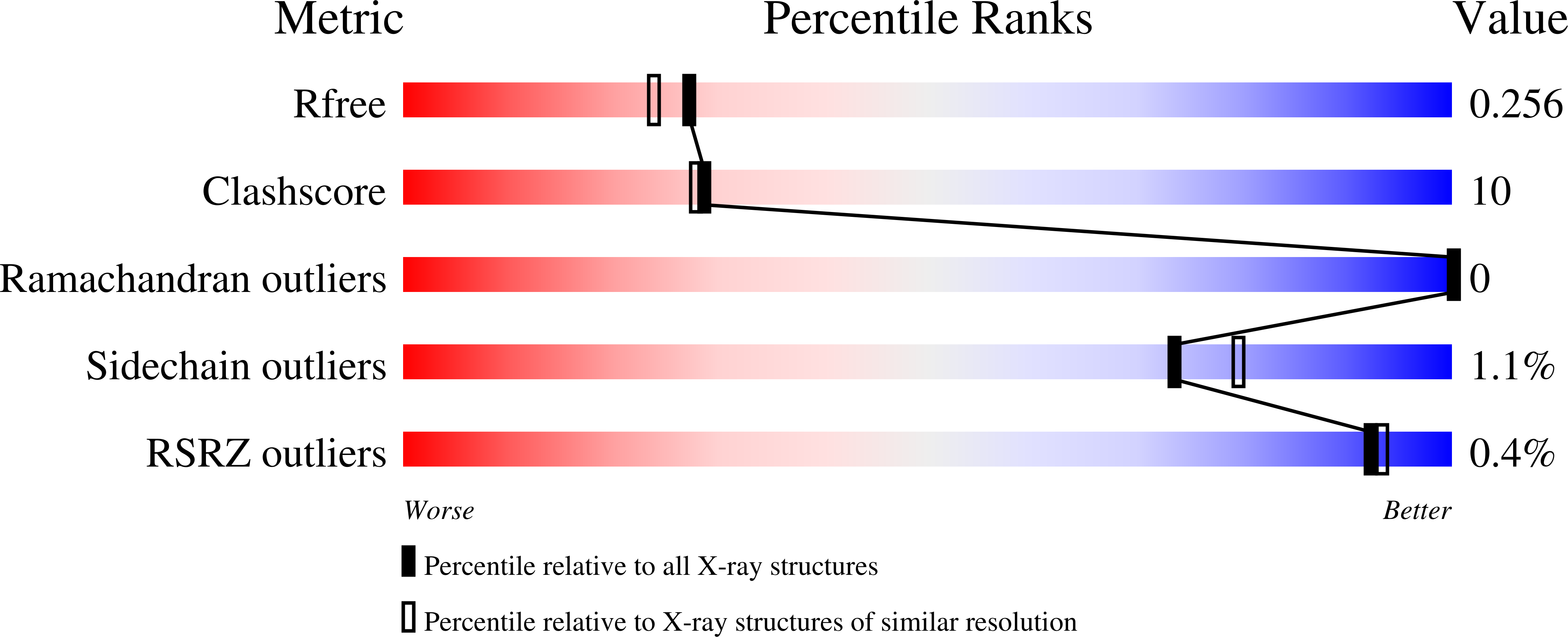
    4WCW

  • PubMed Abstract: 

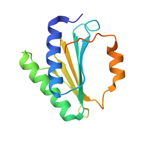
    The ribosomal silencing factor RsfS slows cell growth by inhibiting protein synthesis during periods of diminished nutrient availability. The crystal structure of Mycobacterium tuberculosis (Mtb) RsfS, together with the cryo-electron microscopy (EM) structure of the large subunit 50S of Mtb ribosome, reveals how inhibition of protein synthesis by RsfS occurs. RsfS binds to the 50S at L14, which, when occupied, blocks the association of the small subunit 30S. Although Mtb RsfS is a dimer in solution, only a single subunit binds to 50S. The overlap between the dimer interface and the L14 binding interface confirms that the RsfS dimer must first dissociate to a monomer in order to bind to L14. RsfS interacts primarily through electrostatic and hydrogen bonding to L14. The EM structure shows extended rRNA density that it is not found in the Escherichia coli ribosome, the most striking of these being the extended RNA helix of H54a.


  • Organizational Affiliation

    Department of Biochemistry and Biophysics, Texas A&M University, College Station, TX 77843, USA.


Macromolecules
Find similar proteins by:  (by identity cutoff)  |  3D Structure
Entity ID: 1
MoleculeChains Sequence LengthOrganismDetailsImage
Ribosomal silencing factor RsfS
A, B, C, D
139Mycobacterium tuberculosis H37RvMutation(s): 1 
Gene Names: rsfSRv2420cP425_02518RVBD_2420c
UniProt
Find proteins for O86327 (Mycobacterium tuberculosis (strain ATCC 25618 / H37Rv))
Explore O86327 
Go to UniProtKB:  O86327
Entity Groups  
Sequence Clusters30% Identity50% Identity70% Identity90% Identity95% Identity100% Identity
UniProt GroupO86327
Sequence Annotations
Expand
  • Reference Sequence
Experimental Data & Validation

Experimental Data

  • Method: X-RAY DIFFRACTION
  • Resolution: 2.10 Å
  • R-Value Free: 0.256 
  • R-Value Work: 0.210 
  • R-Value Observed: 0.213 
  • Space Group: P 1
Unit Cell:
Length ( Å )Angle ( ˚ )
a = 50.975α = 110.27
b = 50.975β = 96.17
c = 64.546γ = 110.59
Software Package:
Software NamePurpose
PHENIXrefinement

Structure Validation

View Full Validation Report


Entry History & Funding Information

Deposition Data


Funding OrganizationLocationGrant Number
National Institutes of Health/National Institute Of Allergy and Infectious Diseases (NIH/NIAID)United StatesP01AI095208
Welch FoundationUnited StatesA-0015

Revision History  (Full details and data files)

  • Version 1.0: 2014-09-24
    Type: Initial release
  • Version 1.1: 2015-09-09
    Changes: Database references
  • Version 1.2: 2015-10-21
    Changes: Database references
  • Version 1.3: 2017-09-06
    Changes: Author supporting evidence, Database references, Derived calculations
  • Version 1.4: 2017-11-01
    Changes: Author supporting evidence
  • Version 1.5: 2019-12-11
    Changes: Author supporting evidence
  • Version 1.6: 2023-09-27
    Changes: Data collection, Database references, Derived calculations, Refinement description