4V20

The 3-D structure of the cellobiohydrolase, Cel7A, from Aspergillus fumigatus, disaccharide complex


Experimental Data Snapshot

  • Method: X-RAY DIFFRACTION
  • Resolution: 1.50 Å
  • R-Value Free: 0.158 
  • R-Value Work: 0.132 
  • R-Value Observed: 0.133 

wwPDB Validation   3D Report Full Report


Ligand Structure Quality Assessment 


This is version 3.1 of the entry. See complete history


Literature

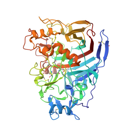
The Three-Dimensional Structure of the Cellobiohydrolase Cel7A from Aspergillus Fumigatus at 1.5 A Resolution

Moroz, O.V.Maranta, M.Shaghasi, T.Harris, P.V.Wilson, K.S.Davies, G.J.

(2015) Acta Crystallogr Sect F Struct Biol Cryst Commun 71: 114

  • DOI: https://doi.org/10.1107/S2053230X14027307
  • Primary Citation of Related Structures:  
    4V1Z, 4V20

  • PubMed Abstract: 

    The enzymatic degradation of plant cell-wall cellulose is central to many industrial processes, including second-generation biofuel production. Key players in this deconstruction are the fungal cellobiohydrolases (CBHs), notably those from family GH7 of the carbohydrate-active enzymes (CAZY) database, which are generally known as CBHI enzymes. Here, three-dimensional structures are reported of the Aspergillus fumigatus CBHI Cel7A solved in uncomplexed and disaccharide-bound forms at resolutions of 1.8 and 1.5 Å, respectively. The product complex with a disaccharide in the +1 and +2 subsites adds to the growing three-dimensional insight into this family of industrially relevant biocatalysts.


  • Organizational Affiliation

    Department of Chemistry, University of York, York Structural Biology Laboratory, York YO10 5DD, England.


Macromolecules
Find similar proteins by:  (by identity cutoff)  |  3D Structure
Entity ID: 1
MoleculeChains Sequence LengthOrganismDetailsImage
CELLOBIOHYDROLASE440Aspergillus fumigatusMutation(s): 0 
EC: 3.2.1.91
UniProt
Find proteins for Q4WM08 (Aspergillus fumigatus (strain ATCC MYA-4609 / CBS 101355 / FGSC A1100 / Af293))
Explore Q4WM08 
Go to UniProtKB:  Q4WM08
Entity Groups  
Sequence Clusters30% Identity50% Identity70% Identity90% Identity95% Identity100% Identity
UniProt GroupQ4WM08
Sequence Annotations
Expand
  • Reference Sequence
Oligosaccharides

Help

Entity ID: 2
MoleculeChains Length2D Diagram Glycosylation3D Interactions
beta-D-glucopyranose-(1-4)-2-deoxy-2-fluoro-beta-D-glucopyranose
B
2N/A
Glycosylation Resources
GlyTouCan:  G28308WE
GlyCosmos:  G28308WE
Biologically Interesting Molecules (External Reference) 1 Unique
Experimental Data & Validation

Experimental Data

  • Method: X-RAY DIFFRACTION
  • Resolution: 1.50 Å
  • R-Value Free: 0.158 
  • R-Value Work: 0.132 
  • R-Value Observed: 0.133 
  • Space Group: P 21 21 2
Unit Cell:
Length ( Å )Angle ( ˚ )
a = 79.56α = 90
b = 130.78β = 90
c = 46.12γ = 90
Software Package:
Software NamePurpose
REFMACrefinement
MOLREPphasing

Structure Validation

View Full Validation Report


Ligand Structure Quality Assessment 


Entry History 

Deposition Data

Revision History  (Full details and data files)

  • Version 1.0: 2015-01-14
    Type: Initial release
  • Version 1.1: 2015-02-04
    Changes: Database references
  • Version 1.2: 2015-02-18
    Changes: Data collection
  • Version 2.0: 2020-03-11
    Changes: Data collection, Derived calculations, Other, Polymer sequence
  • Version 3.0: 2020-07-29
    Type: Remediation
    Reason: Carbohydrate remediation
    Changes: Atomic model, Data collection, Derived calculations, Non-polymer description, Structure summary
  • Version 3.1: 2024-01-10
    Changes: Data collection, Database references, Refinement description, Structure summary