4UZG

Crystal structure of group B streptococcus pilus 2b backbone protein SAK_1440


Experimental Data Snapshot

  • Method: X-RAY DIFFRACTION
  • Resolution: 1.06 Å
  • R-Value Free: 0.142 
  • R-Value Work: 0.125 
  • R-Value Observed: 0.126 

wwPDB Validation   3D Report Full Report


This is version 2.2 of the entry. See complete history


Literature

Structure and assembly of group B streptococcus pilus 2b backbone protein.

Cozzi, R.Malito, E.Lazzarin, M.Nuccitelli, A.Castagnetti, A.Bottomley, M.J.Margarit, I.Maione, D.Rinaudo, C.D.

(2015) PLoS One 10: e0125875-e0125875

  • DOI: https://doi.org/10.1371/journal.pone.0125875
  • Primary Citation of Related Structures:  
    4UZG

  • PubMed Abstract: 

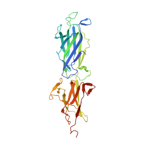
    Group B Streptococcus (GBS) is a major cause of invasive disease in infants. Like other Gram-positive bacteria, GBS uses a sortase C-catalyzed transpeptidation mechanism to generate cell surface pili from backbone and ancillary pilin precursor substrates. The three pilus types identified in GBS contain structural subunits that are highly immunogenic and are promising candidates for the development of a broadly-protective vaccine. Here we report the X-ray crystal structure of the backbone protein of pilus 2b (BP-2b) at 1.06Å resolution. The structure reveals a classical IgG-like fold typical of the pilin subunits of other Gram-positive bacteria. The crystallized portion of the protein (residues 185-468) encompasses domains D2 and D3 that together confer high stability to the protein due to the presence of an internal isopeptide bond within each domain. The D2+D3 region, lacking the N-terminal D1 domain, was as potent as the entire protein in conferring protection against GBS challenge in a well-established mouse model. By site-directed mutagenesis and complementation studies in GBS knock-out strains we identified the residues and motives essential for assembly of the BP-2b monomers into high-molecular weight complexes, thus providing new insights into pilus 2b polymerization.


  • Organizational Affiliation

    Novartis Vaccines and Diagnostics, Siena, Italy.


Macromolecules
Find similar proteins by:  (by identity cutoff)  |  3D Structure
Entity ID: 1
MoleculeChains Sequence LengthOrganismDetailsImage
SURFACE PROTEIN SPB1292Streptococcus agalactiae A909Mutation(s): 0 
UniProt
Find proteins for A0A0M3KKY8 (Streptococcus agalactiae serotype Ia (strain ATCC 27591 / A909 / CDC SS700))
Explore A0A0M3KKY8 
Go to UniProtKB:  A0A0M3KKY8
Entity Groups  
Sequence Clusters30% Identity50% Identity70% Identity90% Identity95% Identity100% Identity
UniProt GroupA0A0M3KKY8
Sequence Annotations
Expand
  • Reference Sequence
Experimental Data & Validation

Experimental Data

  • Method: X-RAY DIFFRACTION
  • Resolution: 1.06 Å
  • R-Value Free: 0.142 
  • R-Value Work: 0.125 
  • R-Value Observed: 0.126 
  • Space Group: P 1 21 1
Unit Cell:
Length ( Å )Angle ( ˚ )
a = 47.64α = 90
b = 53.92β = 90.97
c = 54.77γ = 90
Software Package:
Software NamePurpose
PHENIXrefinement
XDSdata reduction
XSCALEdata scaling
BALBESphasing

Structure Validation

View Full Validation Report


Entry History 

Deposition Data

Revision History  (Full details and data files)

  • Version 1.0: 2015-05-13
    Type: Initial release
  • Version 1.1: 2017-08-09
    Changes: Database references
  • Version 2.0: 2019-10-23
    Changes: Atomic model, Data collection, Derived calculations, Other
  • Version 2.1: 2019-10-30
    Changes: Advisory, Data collection, Derived calculations
  • Version 2.2: 2024-01-10
    Changes: Data collection, Database references, Derived calculations, Refinement description