4UHV

The structure of VgrG1, the needle tip of the bacterial Type VI Secretion System


Experimental Data Snapshot

  • Method: X-RAY DIFFRACTION
  • Resolution: 2.00 Å
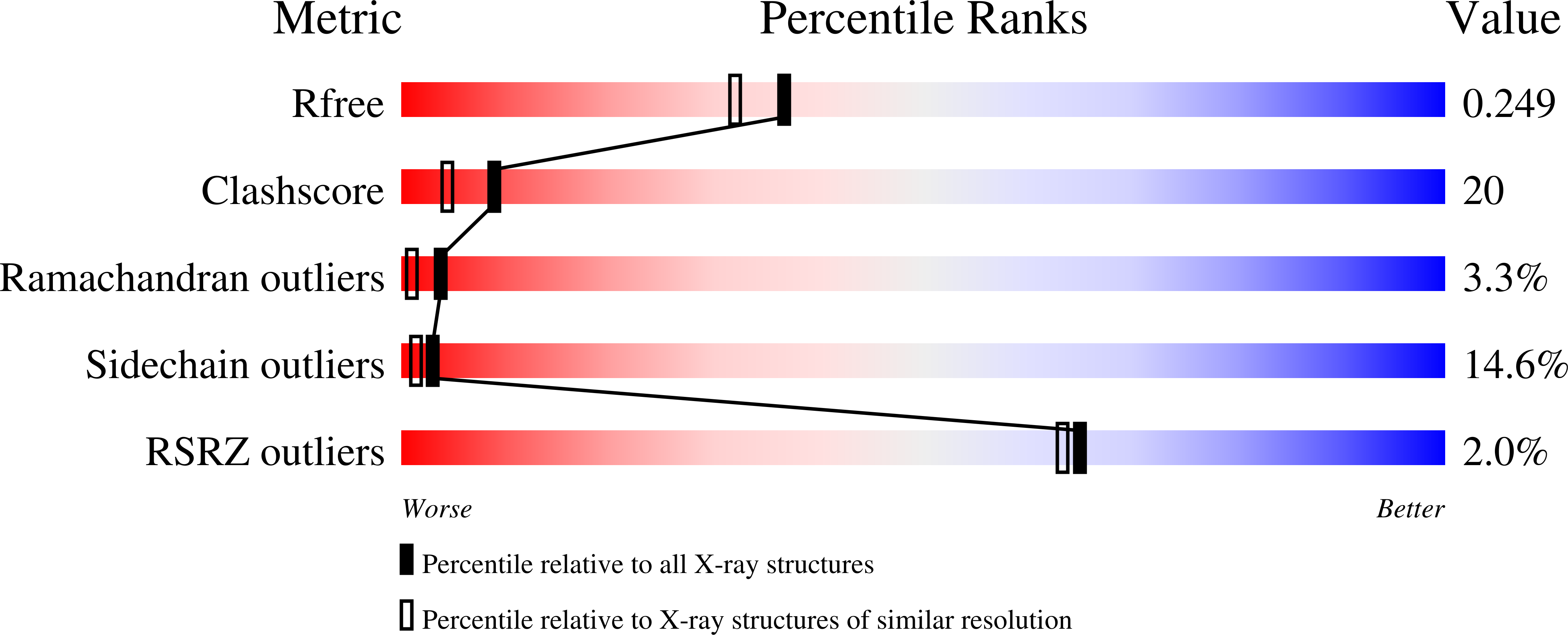
  • R-Value Free: 0.248 
  • R-Value Work: 0.223 
  • R-Value Observed: 0.225 

wwPDB Validation   3D Report Full Report


This is version 1.2 of the entry. See complete history


Literature

The Structure of Vgrg1 from Pseudomonas Aeruginosa, the Needle Tip of the Bacterial Type Vi Secretion System

Spinola-Amilibia, M.Davo-Siguero, I.Ruiz, F.M.Santillana, E.Medrano, F.J.Romero, A.

(2016) Acta Crystallogr D Biol Crystallogr 72: 34

  • DOI: https://doi.org/10.1107/S205979831502149X
  • Primary Citation of Related Structures:  
    4UHV

  • PubMed Abstract: 

    When 300 kV cryo-EM images at Scherzer focus are acquired from ∼ 100 nm thick three-dimensional protein nanocrystals using a Falcon 2 direct electron detector, Fourier transformation can reveal the crystalline lattice to surprisingly high resolutions, even though the images themselves seem to be devoid of any contrast. Here, it is reported how this lattice information can be enhanced by means of a wave finder in combination with Wiener-type maximum-likelihood filtering. This procedure paves the way towards full three-dimensional structure determination at high resolution for protein crystals.


  • Organizational Affiliation

    Biophysical Structural Chemistry, Leiden University, Einsteinweg 55, 2333 CC Leiden, The Netherlands.


Macromolecules
Find similar proteins by:  (by identity cutoff)  |  3D Structure
Entity ID: 1
MoleculeChains Sequence LengthOrganismDetailsImage
VGRG1, VALINE-GLYCINE REPEAT PROTEIN G1
A, B
651Pseudomonas aeruginosa PAO1Mutation(s): 0 
UniProt
Find proteins for Q9I741 (Pseudomonas aeruginosa (strain ATCC 15692 / DSM 22644 / CIP 104116 / JCM 14847 / LMG 12228 / 1C / PRS 101 / PAO1))
Explore Q9I741 
Go to UniProtKB:  Q9I741
Entity Groups  
Sequence Clusters30% Identity50% Identity70% Identity90% Identity95% Identity100% Identity
UniProt GroupQ9I741
Sequence Annotations
Expand
  • Reference Sequence
Small Molecules
Ligands 2 Unique
IDChains Name / Formula / InChI Key2D Diagram3D Interactions
CL
Query on CL

Download Ideal Coordinates CCD File 
AA [auth B]
BA [auth B]
C [auth A]
CA [auth B]
D [auth A]
AA [auth B],
BA [auth B],
C [auth A],
CA [auth B],
D [auth A],
DA [auth B],
E [auth A],
EA [auth B],
F [auth A],
FA [auth B],
G [auth A],
GA [auth B],
H [auth A],
HA [auth B],
I [auth A],
IA [auth B],
JA [auth B],
KA [auth B],
N [auth A],
NA [auth B],
O [auth A],
OA [auth B],
P [auth A],
PA [auth B],
Q [auth A],
QA [auth B],
R [auth A],
RA [auth B],
S [auth A],
SA [auth B],
TA [auth B],
UA [auth B],
V [auth B],
VA [auth B],
W [auth B],
X [auth B],
Y [auth B],
Z [auth B]
CHLORIDE ION
Cl
VEXZGXHMUGYJMC-UHFFFAOYSA-M
NA
Query on NA

Download Ideal Coordinates CCD File 
AB [auth B]
J [auth A]
K [auth A]
L [auth A]
LA [auth B]
AB [auth B],
J [auth A],
K [auth A],
L [auth A],
LA [auth B],
M [auth A],
MA [auth B],
T [auth A],
U [auth A],
WA [auth B],
XA [auth B],
YA [auth B],
ZA [auth B]
SODIUM ION
Na
FKNQFGJONOIPTF-UHFFFAOYSA-N
Modified Residues  1 Unique
IDChains TypeFormula2D DiagramParent
MSE
Query on MSE
A, B
L-PEPTIDE LINKINGC5 H11 N O2 SeMET
Experimental Data & Validation

Experimental Data

  • Method: X-RAY DIFFRACTION
  • Resolution: 2.00 Å
  • R-Value Free: 0.248 
  • R-Value Work: 0.223 
  • R-Value Observed: 0.225 
  • Space Group: P 63
Unit Cell:
Length ( Å )Angle ( ˚ )
a = 78.62α = 90
b = 78.62β = 90
c = 430.32γ = 120
Software Package:
Software NamePurpose
REFMACrefinement
XDSdata reduction
Aimlessdata scaling
PHASERphasing

Structure Validation

View Full Validation Report


Entry History 

Deposition Data

Revision History  (Full details and data files)

  • Version 1.0: 2016-01-27
    Type: Initial release
  • Version 1.1: 2016-03-02
    Changes: Database references
  • Version 1.2: 2024-01-10
    Changes: Data collection, Database references, Derived calculations, Other, Refinement description