4UDM

Crystal structure of Im3 in complex with Y52A mutant of E3RNase


Experimental Data Snapshot

  • Method: X-RAY DIFFRACTION
  • Resolution: 2.96 Å
  • R-Value Free: 0.214 
  • R-Value Work: 0.184 
  • R-Value Observed: 0.186 

wwPDB Validation   3D Report Full Report


This is version 1.1 of the entry. See complete history


Literature

Consequences of Inducing Intrinsic Disorder in a High-Affinity Protein-Protein Interaction.

Papadakos, G.Sharma, A.Lancaster, L.E.Bowen, R.Kaminska, R.Leech, A.P.Walker, D.Redfield, C.Kleanthous, C.

(2015) J Am Chem Soc 137: 5252

  • DOI: https://doi.org/10.1021/ja512607r
  • Primary Citation of Related Structures:  
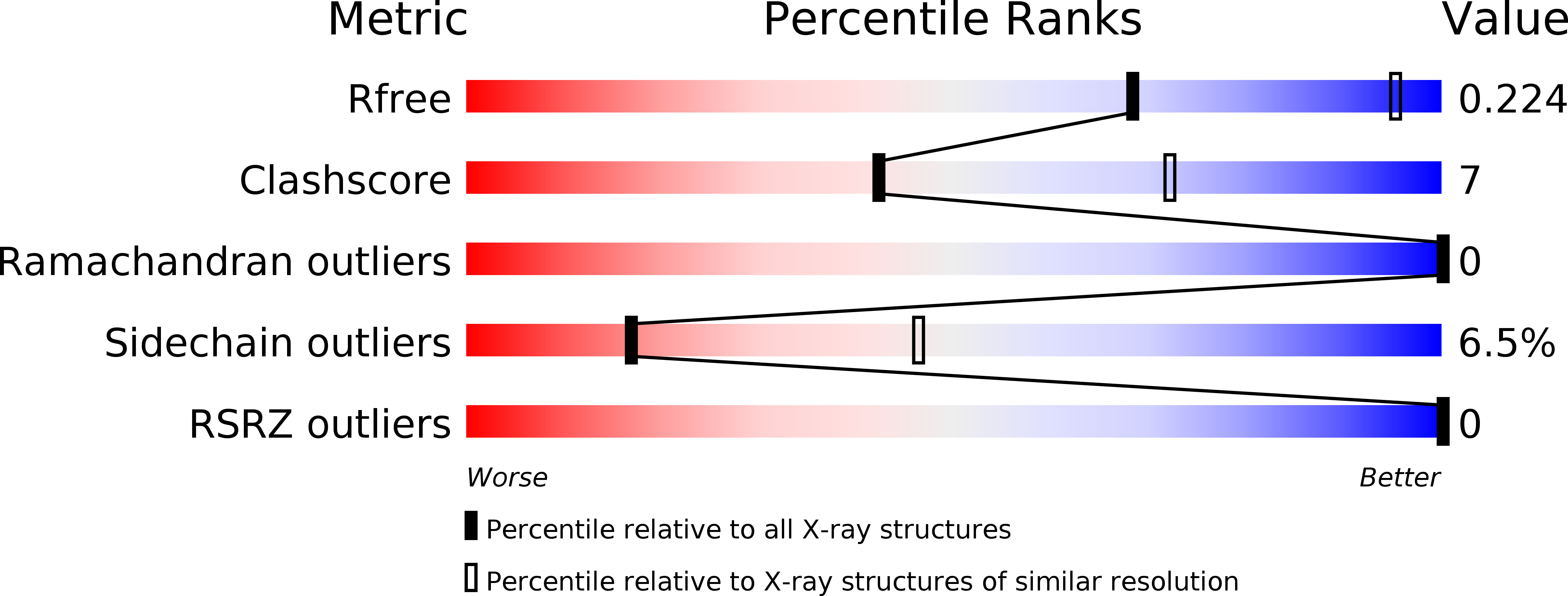
    4UDM

  • PubMed Abstract: 

    The kinetic and thermodynamic consequences of intrinsic disorder in protein-protein recognition are controversial. We address this by inducing one partner of the high-affinity colicin E3 rRNase domain-Im3 complex (K(d) ≈ 10(-12) M) to become an intrinsically disordered protein (IDP). Through a variety of biophysical measurements, we show that a single alanine mutation at Tyr507 within the hydrophobic core of the isolated colicin E3 rRNase domain causes the enzyme to become an IDP (E3 rRNase(IDP)). E3 rRNase(IDP) binds stoichiometrically to Im3 and forms a structure that is essentially identical to the wild-type complex. However, binding of E3 rRNase(IDP) to Im3 is 4 orders of magnitude weaker than that of the folded rRNase, with thermodynamic parameters reflecting the disorder-to-order transition on forming the complex. Critically, pre-steady-state kinetic analysis of the E3 rRNase(IDP)-Im3 complex demonstrates that the decrease in affinity is mostly accounted for by a drop in the electrostatically steered association rate. Our study shows that, notwithstanding the advantages intrinsic disorder brings to biological systems, this can come at severe kinetic and thermodynamic cost.


  • Organizational Affiliation

    †Department of Biochemistry, University of Oxford, South Parks Road, Oxford OX1 3QU, United Kingdom.


Macromolecules
Find similar proteins by:  (by identity cutoff)  |  3D Structure
Entity ID: 1
MoleculeChains Sequence LengthOrganismDetailsImage
COLICIN-E3 IMMUNITY PROTEIN85Escherichia coli BL21(DE3)Mutation(s): 0 
UniProt
Find proteins for P02984 (Escherichia coli)
Explore P02984 
Go to UniProtKB:  P02984
Entity Groups  
Sequence Clusters30% Identity50% Identity70% Identity90% Identity95% Identity100% Identity
UniProt GroupP02984
Sequence Annotations
Expand
  • Reference Sequence
Find similar proteins by:  (by identity cutoff)  |  3D Structure
Entity ID: 2
MoleculeChains Sequence LengthOrganismDetailsImage
COLICIN-E396Escherichia coli BL21(DE3)Mutation(s): 1 
UniProt
Find proteins for P00646 (Escherichia coli)
Explore P00646 
Go to UniProtKB:  P00646
Entity Groups  
Sequence Clusters30% Identity50% Identity70% Identity90% Identity95% Identity100% Identity
UniProt GroupP00646
Sequence Annotations
Expand
  • Reference Sequence
Experimental Data & Validation

Experimental Data

  • Method: X-RAY DIFFRACTION
  • Resolution: 2.96 Å
  • R-Value Free: 0.214 
  • R-Value Work: 0.184 
  • R-Value Observed: 0.186 
  • Space Group: P 31 2 1
Unit Cell:
Length ( Å )Angle ( ˚ )
a = 92.854α = 90
b = 92.854β = 90
c = 76.942γ = 120
Software Package:
Software NamePurpose
REFMACrefinement
HKL-2000data reduction
HKL-2000data scaling
MOLREPphasing

Structure Validation

View Full Validation Report


Entry History 

Deposition Data

Revision History  (Full details and data files)

  • Version 1.0: 2016-01-13
    Type: Initial release
  • Version 1.1: 2023-12-20
    Changes: Data collection, Database references, Derived calculations, Other, Refinement description