4UDG

Crystal structure of b-1,4-mannopyranosyl-chitobiose phosphorylase at 1.60 Angstrom in complex with N-acetylglucosamine and inorganic phosphate


Experimental Data Snapshot

  • Method: X-RAY DIFFRACTION
  • Resolution: 1.60 Å
  • R-Value Free: 0.183 
  • R-Value Work: 0.154 
  • R-Value Observed: 0.155 

wwPDB Validation   3D Report Full Report


Ligand Structure Quality Assessment 


This is version 2.1 of the entry. See complete history


Literature

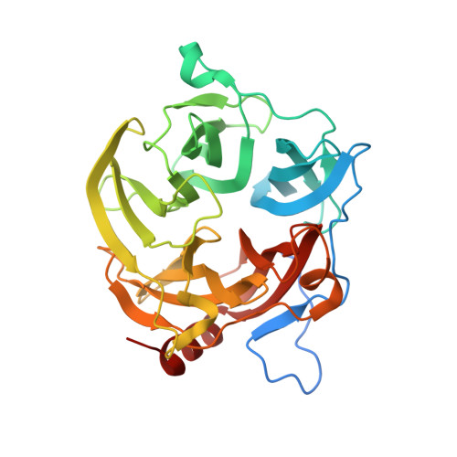
Structural Bases for N-Glycan Processing by Mannoside Phosphorylase.

Ladeveze, S.Cioci, G.Roblin, P.Mourey, L.Tranier, S.Potocki-Veronese, G.

(2015) Acta Crystallogr D Biol Crystallogr 71: 1335

  • DOI: https://doi.org/10.1107/S1399004715006604
  • Primary Citation of Related Structures:  
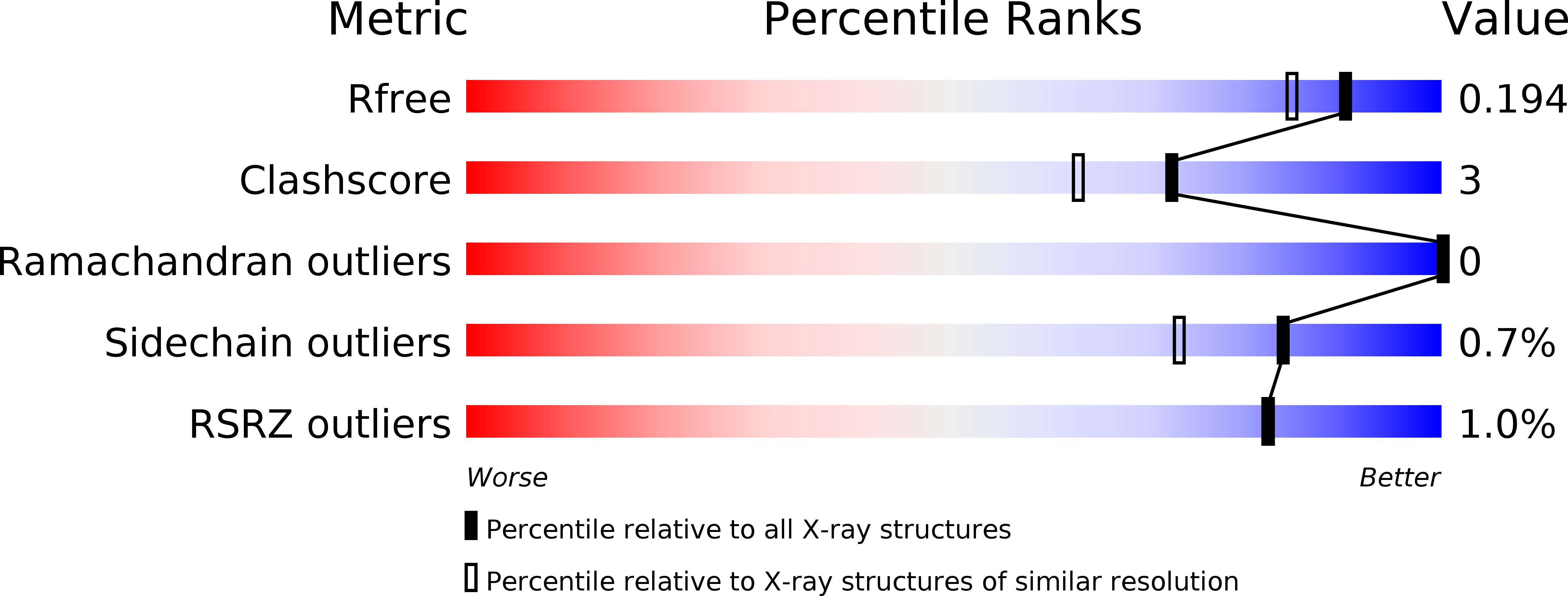
    4UDG, 4UDI, 4UDJ, 4UDK

  • PubMed Abstract: 

    The first crystal structure of Uhgb_MP, a β-1,4-mannopyranosyl-chitobiose phosphorylase belonging to the GH130 family which is involved in N-glycan degradation by human gut bacteria, was solved at 1.85 Å resolution in the apo form and in complex with mannose and N-acetylglucosamine. SAXS and crystal structure analysis revealed a hexameric structure, a specific feature of GH130 enzymes among other glycoside phosphorylases. Mapping of the -1 and +1 subsites in the presence of phosphate confirmed the conserved Asp104 as the general acid/base catalytic residue, which is in agreement with a single-step reaction mechanism involving Man O3 assistance for proton transfer. Analysis of this structure, the first to be solved for a member of the GH130_2 subfamily, revealed Met67, Phe203 and the Gly121-Pro125 loop as the main determinants of the specificity of Uhgb_MP and its homologues towards the N-glycan core oligosaccharides and mannan, and the molecular bases of the key role played by GH130 enzymes in the catabolism of dietary fibre and host glycans.


  • Organizational Affiliation

    Université de Toulouse; INSA, UPS, INP; LISBP, 135 Avenue de Rangueil, 31077 Toulouse, France.


Macromolecules
Find similar proteins by:  (by identity cutoff)  |  3D Structure
Entity ID: 1
MoleculeChains Sequence LengthOrganismDetailsImage
UHGB_MP
A, B, C, D, E
A, B, C, D, E, F
347uncultured organismMutation(s): 0 
EC: 2.4.1
UniProt
Find proteins for D9ZDQ9 (uncultured organism)
Explore D9ZDQ9 
Go to UniProtKB:  D9ZDQ9
Entity Groups  
Sequence Clusters30% Identity50% Identity70% Identity90% Identity95% Identity100% Identity
UniProt GroupD9ZDQ9
Sequence Annotations
Expand
  • Reference Sequence
Small Molecules
Ligands 5 Unique
IDChains Name / Formula / InChI Key2D Diagram3D Interactions
NDG
Query on NDG

Download Ideal Coordinates CCD File 
AA [auth E]
GA [auth F]
H [auth A]
N [auth B]
S [auth C]
AA [auth E],
GA [auth F],
H [auth A],
N [auth B],
S [auth C],
W [auth D]
2-acetamido-2-deoxy-alpha-D-glucopyranose
C8 H15 N O6
OVRNDRQMDRJTHS-PVFLNQBWSA-N
PO4
Query on PO4

Download Ideal Coordinates CCD File 
FA [auth F]
G [auth A]
M [auth B]
R [auth C]
V [auth D]
FA [auth F],
G [auth A],
M [auth B],
R [auth C],
V [auth D],
Z [auth E]
PHOSPHATE ION
O4 P
NBIIXXVUZAFLBC-UHFFFAOYSA-K
GOL
Query on GOL

Download Ideal Coordinates CCD File 
BA [auth E]
HA [auth F]
I [auth A]
O [auth B]
P [auth B]
BA [auth E],
HA [auth F],
I [auth A],
O [auth B],
P [auth B],
T [auth C],
X [auth D]
GLYCEROL
C3 H8 O3
PEDCQBHIVMGVHV-UHFFFAOYSA-N
EDO
Query on EDO

Download Ideal Coordinates CCD File 
DA [auth E],
EA [auth E],
JA [auth F],
K [auth A],
L [auth A]
1,2-ETHANEDIOL
C2 H6 O2
LYCAIKOWRPUZTN-UHFFFAOYSA-N
K
Query on K

Download Ideal Coordinates CCD File 
CA [auth E]
IA [auth F]
J [auth A]
Q [auth B]
U [auth C]
CA [auth E],
IA [auth F],
J [auth A],
Q [auth B],
U [auth C],
Y [auth D]
POTASSIUM ION
K
NPYPAHLBTDXSSS-UHFFFAOYSA-N
Experimental Data & Validation

Experimental Data

  • Method: X-RAY DIFFRACTION
  • Resolution: 1.60 Å
  • R-Value Free: 0.183 
  • R-Value Work: 0.154 
  • R-Value Observed: 0.155 
  • Space Group: P 21 21 21
Unit Cell:
Length ( Å )Angle ( ˚ )
a = 83.816α = 90
b = 140.912β = 90
c = 168.598γ = 90
Software Package:
Software NamePurpose
REFMACrefinement
XDSdata reduction
XDSdata scaling
PHASERphasing

Structure Validation

View Full Validation Report


Ligand Structure Quality Assessment 


Entry History 

Deposition Data

Revision History  (Full details and data files)

  • Version 1.0: 2015-05-27
    Type: Initial release
  • Version 1.1: 2015-06-17
    Changes: Database references
  • Version 2.0: 2020-07-29
    Type: Remediation
    Reason: Carbohydrate remediation
    Changes: Atomic model, Data collection, Derived calculations, Other, Structure summary
  • Version 2.1: 2023-12-20
    Changes: Data collection, Database references, Refinement description, Structure summary