4TXI

Construct of MICAL-1 containing the monooxygenase and calponin homology domains


Experimental Data Snapshot

  • Method: X-RAY DIFFRACTION
  • Resolution: 2.31 Å
  • R-Value Free: 0.279 
  • R-Value Work: 0.216 
  • R-Value Observed: 0.219 

wwPDB Validation   3D Report Full Report


Ligand Structure Quality Assessment 


This is version 1.4 of the entry. See complete history


Literature

Modulation of MICAL Monooxygenase Activity by its Calponin Homology Domain: Structural and Mechanistic Insights.

Alqassim, S.S.Urquiza, M.Borgnia, E.Nagib, M.Amzel, L.M.Bianchet, M.A.

(2016) Sci Rep 6: 22176-22176

  • DOI: https://doi.org/10.1038/srep22176
  • Primary Citation of Related Structures:  
    4TXI

  • PubMed Abstract: 

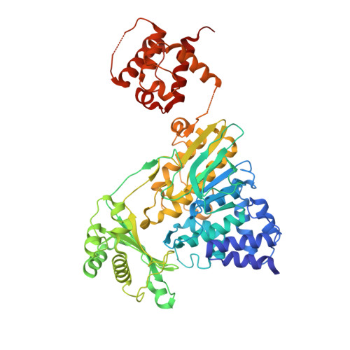
    MICALs (Molecule Interacting with CasL) are conserved multidomain enzymes essential for cytoskeletal reorganization in nerve development, endocytosis, and apoptosis. In these enzymes, a type-2 calponin homology (CH) domain always follows an N-terminal monooxygenase (MO) domain. Although the CH domain is required for MICAL-1 cellular localization and actin-associated function, its contribution to the modulation of MICAL activity towards actin remains unclear. Here, we present the structure of a fragment of MICAL-1 containing the MO and the CH domains-determined by X-ray crystallography and small angle scattering-as well as kinetics experiments designed to probe the contribution of the CH domain to the actin-modification activity. Our results suggest that the CH domain, which is loosely connected to the MO domain by a flexible linker and is far away from the catalytic site, couples F-actin to the enhancement of redox activity of MICALMO-CH by a cooperative mechanism involving a trans interaction between adjacently bound molecules. Binding cooperativity is also observed in other proteins regulating actin assembly/disassembly dynamics, such as ADF/Cofilins.


  • Organizational Affiliation

    Structural Enzymology and Thermodynamics Group, Department of Biophysics &Biophysical Chemistry, Johns Hopkins University School of Medicine, 725 North Wolfe St., Baltimore, MD 21205, USA.


Macromolecules
Find similar proteins by:  (by identity cutoff)  |  3D Structure
Entity ID: 1
MoleculeChains Sequence LengthOrganismDetailsImage
Protein-methionine sulfoxide oxidase MICAL1615Mus musculusMutation(s): 0 
Gene Names: Mical1MicalNical
EC: 1.14.13
UniProt
Find proteins for Q8VDP3 (Mus musculus)
Explore Q8VDP3 
Go to UniProtKB:  Q8VDP3
Entity Groups  
Sequence Clusters30% Identity50% Identity70% Identity90% Identity95% Identity100% Identity
UniProt GroupQ8VDP3
Sequence Annotations
Expand
  • Reference Sequence
Experimental Data & Validation

Experimental Data

  • Method: X-RAY DIFFRACTION
  • Resolution: 2.31 Å
  • R-Value Free: 0.279 
  • R-Value Work: 0.216 
  • R-Value Observed: 0.219 
  • Space Group: P 1 21 1
Unit Cell:
Length ( Å )Angle ( ˚ )
a = 71.924α = 90
b = 49.885β = 97.03
c = 95.852γ = 90
Software Package:
Software NamePurpose
PHENIXrefinement
HKL-2000data reduction
MOLREPphasing
PDB_EXTRACTdata extraction
HKL-2000data scaling
DENZOdata reduction
SCALEPACKdata scaling

Structure Validation

View Full Validation Report


Ligand Structure Quality Assessment 


Entry History 

Deposition Data

Revision History  (Full details and data files)

  • Version 1.0: 2016-01-13
    Type: Initial release
  • Version 1.1: 2016-07-27
    Changes: Data collection
  • Version 1.2: 2017-08-16
    Changes: Database references, Derived calculations
  • Version 1.3: 2017-11-22
    Changes: Refinement description
  • Version 1.4: 2023-12-27
    Changes: Data collection, Database references