


Crystal structure of Dihydropyrimidinase from Brucella suis
Seattle Structural Genomics Center for Infectious Disease (SSGCID), Abendroth, J., Davies, D.R., Lorimer, D., Edwards, T.E.To be published.
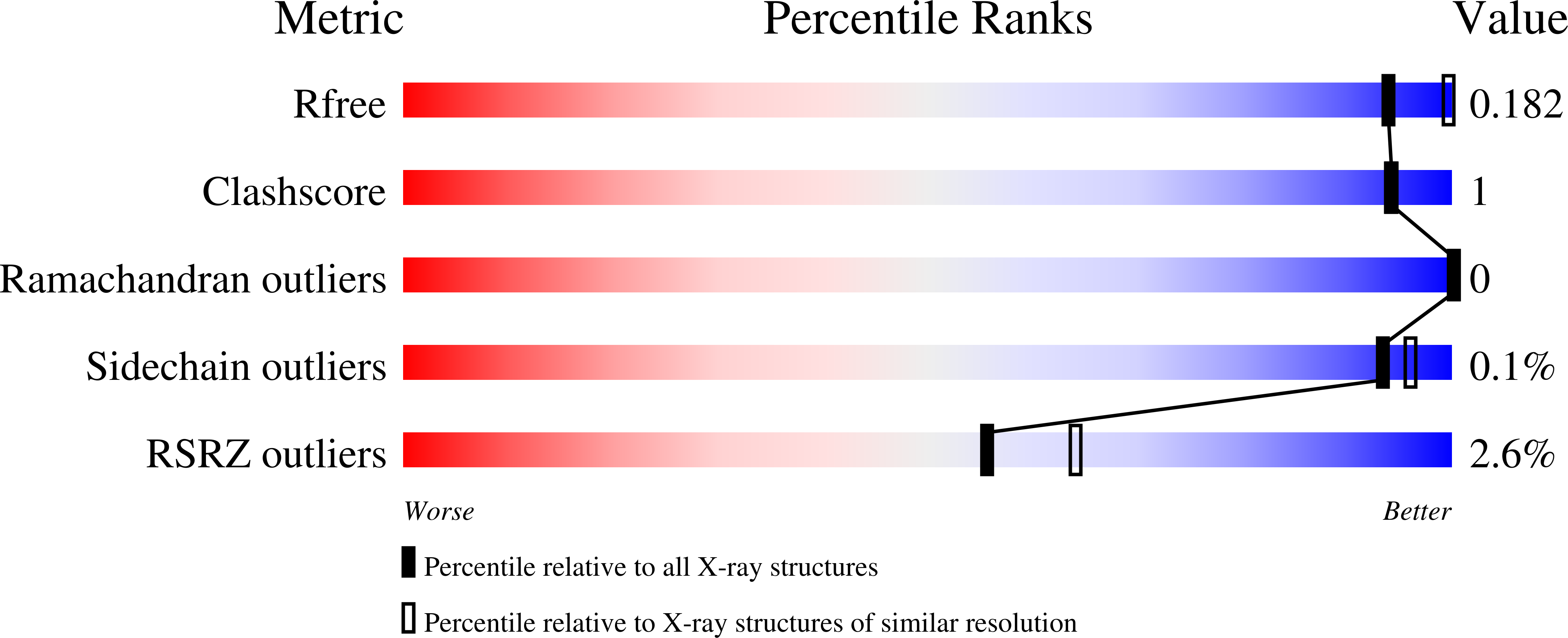
Experimental Data Snapshot
wwPDB Validation 3D Report Full Report
Entity ID: 1 | |||||
|---|---|---|---|---|---|
| Molecule | Chains | Sequence Length | Organism | Details | Image |
| D-hydantoinase | 497 | Brucella suis 1330 | Mutation(s): 0 Gene Names: dhT, BR0278, BS1330_I0279 EC: 3.5.2.2 | 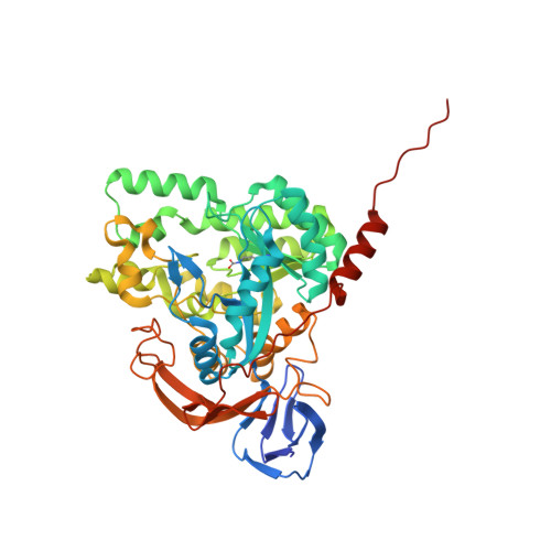 | |
UniProt | |||||
Find proteins for A0A0H3G9X2 (Brucella suis biovar 1 (strain 1330)) Explore A0A0H3G9X2 Go to UniProtKB: A0A0H3G9X2 | |||||
Entity Groups | |||||
| Sequence Clusters | 30% Identity50% Identity70% Identity90% Identity95% Identity100% Identity | ||||
| UniProt Group | A0A0H3G9X2 | ||||
Sequence AnnotationsExpand | |||||
| |||||
| Ligands 2 Unique | |||||
|---|---|---|---|---|---|
| ID | Chains | Name / Formula / InChI Key | 2D Diagram | 3D Interactions | |
| ZN Query on ZN | EA [auth E] FA [auth E] G [auth A] H [auth A] KA [auth F] | ZINC ION Zn PTFCDOFLOPIGGS-UHFFFAOYSA-N |  | ||
| EDO Query on EDO | AA [auth D] BA [auth D] CA [auth D] DA [auth D] GA [auth E] | 1,2-ETHANEDIOL C2 H6 O2 LYCAIKOWRPUZTN-UHFFFAOYSA-N |  | ||
| Modified Residues 1 Unique | |||||
|---|---|---|---|---|---|
| ID | Chains | Type | Formula | 2D Diagram | Parent |
| KCX Query on KCX | A, B, C, D, E A, B, C, D, E, F | L-PEPTIDE LINKING | C7 H14 N2 O4 |  | LYS |
| Length ( Å ) | Angle ( ˚ ) |
|---|---|
| a = 156.69 | α = 90 |
| b = 88.83 | β = 91.17 |
| c = 221.24 | γ = 90 |
| Software Name | Purpose |
|---|---|
| XDS | data reduction |
| XSCALE | data scaling |
| PHASER | phasing |
| ARP | model building |
| PHENIX | refinement |
| PDB_EXTRACT | data extraction |
RCSB PDB (citation) is hosted by
RCSB PDB is a member of the

