


Crystal structure of the Phosphorybosylpyrophosphate synthetase from E. Coli
Timofeev, V.I., Abramchik, Y.A., Muravieva, T.I., Iaroslavtceva, A.K., Stepanenko, V.N., Zhukhlistova, N.E., Esipov, R.S., Kuranova, I.P.To be published.
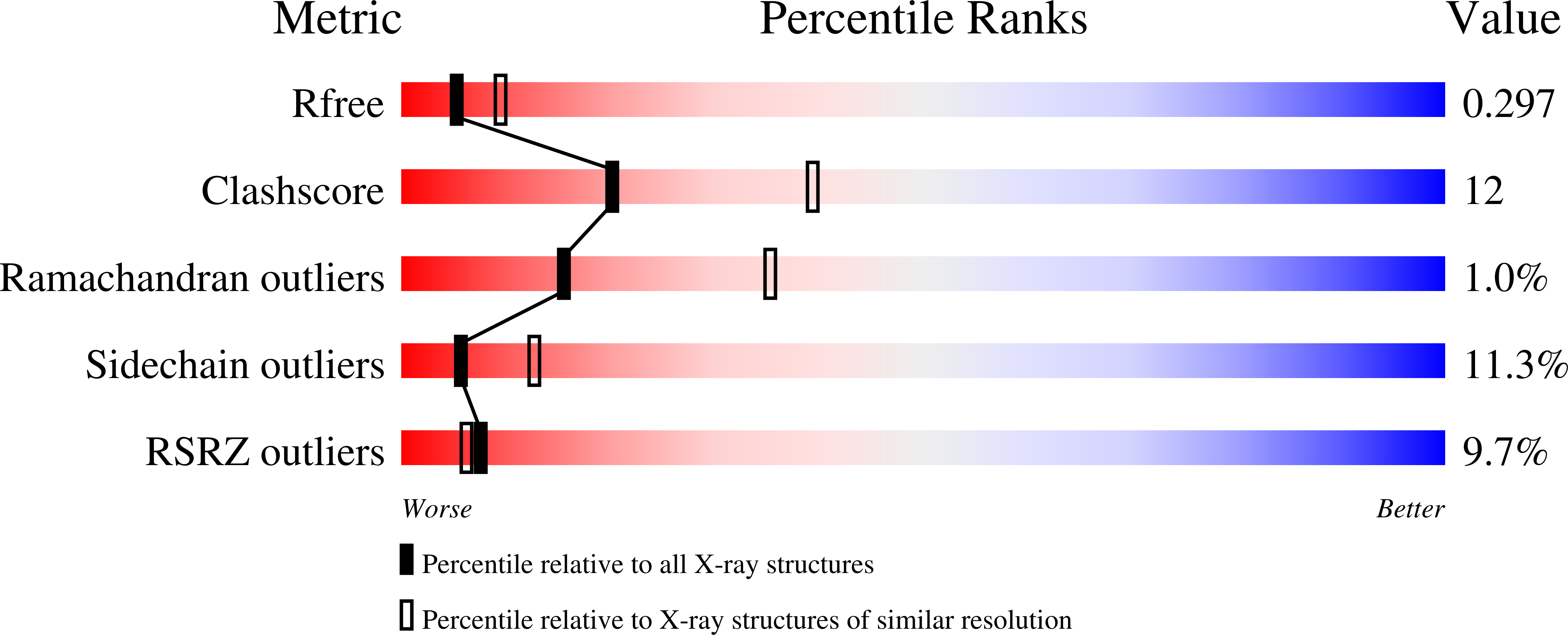
Experimental Data Snapshot
wwPDB Validation 3D Report Full Report
Entity ID: 1 | |||||
|---|---|---|---|---|---|
| Molecule | Chains | Sequence Length | Organism | Details | Image |
| Ribose-phosphate pyrophosphokinase | 316 | Escherichia coli | Mutation(s): 0 Gene Names: prsA, prs EC: 2.7.6.1 | 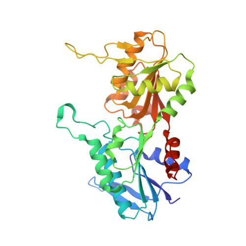 | |
UniProt | |||||
Find proteins for P0A717 (Escherichia coli (strain K12)) Explore P0A717 Go to UniProtKB: P0A717 | |||||
Entity Groups | |||||
| Sequence Clusters | 30% Identity50% Identity70% Identity90% Identity95% Identity100% Identity | ||||
| UniProt Group | P0A717 | ||||
Sequence AnnotationsExpand | |||||
| |||||
| Ligands 1 Unique | |||||
|---|---|---|---|---|---|
| ID | Chains | Name / Formula / InChI Key | 2D Diagram | 3D Interactions | |
| MG Query on MG | B [auth A] | MAGNESIUM ION Mg JLVVSXFLKOJNIY-UHFFFAOYSA-N |  | ||
| Length ( Å ) | Angle ( ˚ ) |
|---|---|
| a = 104.599 | α = 90 |
| b = 104.599 | β = 90 |
| c = 125.78 | γ = 120 |
| Software Name | Purpose |
|---|---|
| HKL-2000 | data collection |
| PHASER | phasing |
| REFMAC | refinement |
| MOSFLM | data reduction |
| SCALA | data scaling |
RCSB PDB (citation) is hosted by
RCSB PDB is a member of the

