4RXA

Crystal structure of human farnesyl diphosphate synthase in complex with BPH-1358


Experimental Data Snapshot

  • Method: X-RAY DIFFRACTION
  • Resolution: 2.20 Å
  • R-Value Free: 0.263 
  • R-Value Work: 0.221 
  • R-Value Observed: 0.223 

wwPDB Validation   3D Report Full Report


Ligand Structure Quality Assessment 


This is version 1.1 of the entry. See complete history


Literature

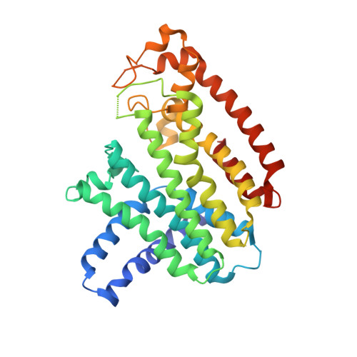
Farnesyl diphosphate synthase inhibitors with unique ligand-binding geometries.

Liu, Y.L.Cao, R.Wang, Y.Oldfield, E.

(2015) ACS Med Chem Lett 6: 349-354

  • DOI: https://doi.org/10.1021/ml500528x
  • Primary Citation of Related Structures:  
    4RXA, 4RXC, 4RXD, 4RXE, 4RYP

  • PubMed Abstract: 

    Farnesyl diphosphate synthase (FPPS) is an important drug target for bone resorption, cancer, and some infectious diseases. Here, we report five new structures including two having unique bound ligand geometries. The diamidine inhibitor 7 binds to human FPPS close to the homoallylic (S2) and allosteric (S3) sites and extends into a new site, here called S4. With the bisphosphonate inhibitor 8, two molecules bind to Trypanosoma brucei FPPS, one molecule in the allylic site (S1) and the other close to S2, the first observation of two bisphosphonate molecules bound to FPPS. We also report the structures of apo-FPPS from T. brucei, together with two more bisphosphonate-bound structures (2,9), for purposes of comparison. The diamidine structure is of particular interest because 7 could represent a new lead for lipophilic FPPS inhibitors, while 8 has low micromolar activity against T. brucei, the causative agent of human African trypanosomiasis.


  • Organizational Affiliation

    Department of Chemistry, University of Illinois at Urbana-Champaign , Urbana, Illinois 61801, United States.


Macromolecules
Find similar proteins by:  (by identity cutoff)  |  3D Structure
Entity ID: 1
MoleculeChains Sequence LengthOrganismDetailsImage
Farnesyl pyrophosphate synthase348Homo sapiensMutation(s): 0 
Gene Names: FDPSFPSKIAA1293
EC: 2.5.1.10 (PDB Primary Data), 2.5.1.1 (PDB Primary Data)
UniProt & NIH Common Fund Data Resources
Find proteins for P14324 (Homo sapiens)
Explore P14324 
Go to UniProtKB:  P14324
PHAROS:  P14324
GTEx:  ENSG00000160752 
Entity Groups  
Sequence Clusters30% Identity50% Identity70% Identity90% Identity95% Identity100% Identity
UniProt GroupP14324
Sequence Annotations
Expand
  • Reference Sequence
Small Molecules
Ligands 2 Unique
IDChains Name / Formula / InChI Key2D Diagram3D Interactions
3F2
Query on 3F2

Download Ideal Coordinates CCD File 
B [auth A]N,N'-bis[3-(4,5-dihydro-1H-imidazol-2-yl)phenyl]biphenyl-4,4'-dicarboxamide
C32 H28 N6 O2
HOYOJUMMRGKESB-UHFFFAOYSA-N
PO4
Query on PO4

Download Ideal Coordinates CCD File 
C [auth A]PHOSPHATE ION
O4 P
NBIIXXVUZAFLBC-UHFFFAOYSA-K
Binding Affinity Annotations 
IDSourceBinding Affinity
3F2 Binding MOAD:  4RXA IC50: 1800 (nM) from 1 assay(s)
BindingDB:  4RXA IC50: 1800 (nM) from 1 assay(s)
Experimental Data & Validation

Experimental Data

  • Method: X-RAY DIFFRACTION
  • Resolution: 2.20 Å
  • R-Value Free: 0.263 
  • R-Value Work: 0.221 
  • R-Value Observed: 0.223 
  • Space Group: P 41 21 2
Unit Cell:
Length ( Å )Angle ( ˚ )
a = 110.757α = 90
b = 110.757β = 90
c = 77.96γ = 90
Software Package:
Software NamePurpose
REFMACrefinement
PDB_EXTRACTdata extraction
HKL-3000data collection
HKL-3000data reduction
HKL-3000data scaling
PHASESphasing

Structure Validation

View Full Validation Report


Ligand Structure Quality Assessment 


Entry History 

Deposition Data

Revision History  (Full details and data files)

  • Version 1.0: 2015-04-15
    Type: Initial release
  • Version 1.1: 2024-02-28
    Changes: Data collection, Database references, Derived calculations