4RNH

PaMorA tandem diguanylate cyclase - phosphodiesterase, c-di-GMP complex


Experimental Data Snapshot

  • Method: X-RAY DIFFRACTION
  • Resolution: 2.45 Å
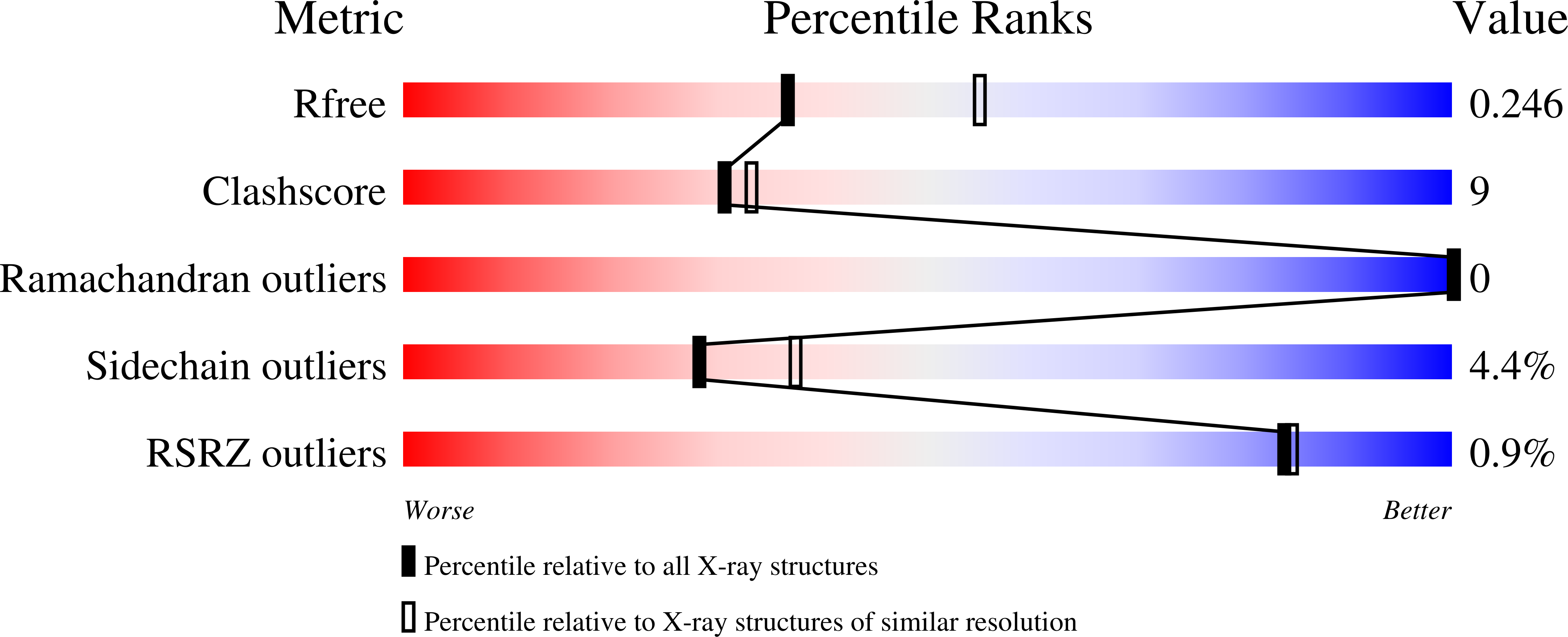
  • R-Value Free: 0.243 
  • R-Value Work: 0.184 
  • R-Value Observed: 0.187 

wwPDB Validation   3D Report Full Report


Ligand Structure Quality Assessment 


This is version 1.3 of the entry. See complete history


Literature

Formation and dimerization of the phosphodiesterase active site of the Pseudomonas aeruginosa MorA, a bi-functional c-di-GMP regulator.

Phippen, C.W.Mikolajek, H.Schlaefli, H.G.Keevil, C.W.Webb, J.S.Tews, I.

(2014) FEBS Lett 588: 4631-4636

  • DOI: https://doi.org/10.1016/j.febslet.2014.11.002
  • Primary Citation of Related Structures:  
    4RNF, 4RNH, 4RNI, 4RNJ

  • PubMed Abstract: 

    Diguanylate cyclases (DGC) and phosphodiesterases (PDE), respectively synthesise and hydrolyse the secondary messenger cyclic dimeric GMP (c-di-GMP), and both activities are often found in a single protein. Intracellular c-di-GMP levels in turn regulate bacterial motility, virulence and biofilm formation. We report the first structure of a tandem DGC-PDE fragment, in which the catalytic domains are shown to be active. Two phosphodiesterase states are distinguished by active site formation. The structures, in the presence or absence of c-di-GMP, suggest that dimerisation and binding pocket formation are linked, with dimerisation being required for catalytic activity. An understanding of PDE activation is important, as biofilm dispersal via c-di-GMP hydrolysis has therapeutic effects on chronic infections.


  • Organizational Affiliation

    Centre for Biological Sciences and Institute for Life Sciences, Life Sciences Building B85, The University of Southampton, University Rd, Southampton, Hampshire, SO17 1BJ, United Kingdom.


Macromolecules
Find similar proteins by:  (by identity cutoff)  |  3D Structure
Entity ID: 1
MoleculeChains Sequence LengthOrganismDetailsImage
Motility regulator453Pseudomonas aeruginosa PAO1Mutation(s): 0 
Gene Names: morAPA4601
EC: 2.7.7.65 (PDB Primary Data), 3.1.4.52 (PDB Primary Data)
UniProt
Find proteins for Q9HVI8 (Pseudomonas aeruginosa (strain ATCC 15692 / DSM 22644 / CIP 104116 / JCM 14847 / LMG 12228 / 1C / PRS 101 / PAO1))
Explore Q9HVI8 
Go to UniProtKB:  Q9HVI8
Entity Groups  
Sequence Clusters30% Identity50% Identity70% Identity90% Identity95% Identity100% Identity
UniProt GroupQ9HVI8
Sequence Annotations
Expand
  • Reference Sequence
Small Molecules
Ligands 2 Unique
IDChains Name / Formula / InChI Key2D Diagram3D Interactions
C2E
Query on C2E

Download Ideal Coordinates CCD File 
D [auth A]9,9'-[(2R,3R,3aS,5S,7aR,9R,10R,10aS,12S,14aR)-3,5,10,12-tetrahydroxy-5,12-dioxidooctahydro-2H,7H-difuro[3,2-d:3',2'-j][1,3,7,9,2,8]tetraoxadiphosphacyclododecine-2,9-diyl]bis(2-amino-1,9-dihydro-6H-purin-6-one)
C20 H24 N10 O14 P2
PKFDLKSEZWEFGL-MHARETSRSA-N
MG
Query on MG

Download Ideal Coordinates CCD File 
B [auth A],
C [auth A]
MAGNESIUM ION
Mg
JLVVSXFLKOJNIY-UHFFFAOYSA-N
Experimental Data & Validation

Experimental Data

  • Method: X-RAY DIFFRACTION
  • Resolution: 2.45 Å
  • R-Value Free: 0.243 
  • R-Value Work: 0.184 
  • R-Value Observed: 0.187 
  • Space Group: P 31 2 1
Unit Cell:
Length ( Å )Angle ( ˚ )
a = 81.3α = 90
b = 81.3β = 90
c = 128.1γ = 120
Software Package:
Software NamePurpose
xia2data reduction
REFMACrefinement
PDB_EXTRACTdata extraction
XDSdata scaling
XDSdata reduction
xia2data scaling
MOLREPphasing

Structure Validation

View Full Validation Report


Ligand Structure Quality Assessment 


Entry History 

Deposition Data

Revision History  (Full details and data files)

  • Version 1.0: 2014-11-19
    Type: Initial release
  • Version 1.1: 2015-02-25
    Changes: Structure summary
  • Version 1.2: 2015-04-22
    Changes: Database references
  • Version 1.3: 2024-02-28
    Changes: Data collection, Database references, Derived calculations, Structure summary