4R9T

L-ficolin complexed to sulphates


Experimental Data Snapshot

  • Method: X-RAY DIFFRACTION
  • Resolution: 2.25 Å
  • R-Value Free: 0.218 
  • R-Value Work: 0.188 
  • R-Value Observed: 0.189 

wwPDB Validation   3D Report Full Report


This is version 2.0 of the entry. See complete history


Literature

Human ficolin-2 recognition versatility extended: An update on the binding of ficolin-2 to sulfated/phosphated carbohydrates.

Laffly, E.Lacroix, M.Martin, L.Vassal-Stermann, E.Thielens, N.M.Gaboriaud, C.

(2014) FEBS Lett 588: 4694-4700

  • DOI: https://doi.org/10.1016/j.febslet.2014.10.042
  • Primary Citation of Related Structures:  
    4R9J, 4R9T

  • PubMed Abstract: 

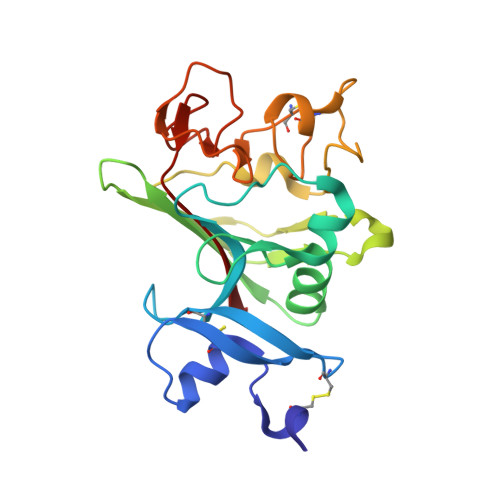
    Ficolin-2 has been reported to bind to DNA and heparin, but the mechanism involved has not been thoroughly investigated. X-ray studies of the ficolin-2 fibrinogen-like domain in complex with several new ligands now show that sulfate and phosphate groups are prone to bind to the S3 binding site of the protein. Composed of Arg132, Asp133, Thr136 and Lys221, the S3 site was previously shown to mainly bind N-acetyl groups. Furthermore, DNA and heparin compete for binding to ficolin-2. Mutagenesis studies reveal that Arg132, and to a lesser extent Asp133, are important for this binding property. The versatility of the S3 site in binding N-acetyl, sulfate and phosphate groups is discussed through comparisons with homologous fibrinogen-like recognition proteins.


  • Organizational Affiliation

    Univ. Grenoble Alpes, IBS, F-38044 Grenoble, France; CNRS, IBS, F-38044 Grenoble, France; CEA, IBS, F-38044 Grenoble, France.


Macromolecules
Find similar proteins by:  (by identity cutoff)  |  3D Structure
Entity ID: 1
MoleculeChains Sequence LengthOrganismDetailsImage
Ficolin-2A [auth B],
B [auth A],
C
218Homo sapiensMutation(s): 0 
Gene Names: FCN2FCNL
UniProt & NIH Common Fund Data Resources
Find proteins for Q15485 (Homo sapiens)
Explore Q15485 
Go to UniProtKB:  Q15485
PHAROS:  Q15485
GTEx:  ENSG00000160339 
Entity Groups  
Sequence Clusters30% Identity50% Identity70% Identity90% Identity95% Identity100% Identity
UniProt GroupQ15485
Sequence Annotations
Expand
  • Reference Sequence
Oligosaccharides

Help

Entity ID: 2
MoleculeChains Length2D Diagram Glycosylation3D Interactions
2-acetamido-2-deoxy-beta-D-glucopyranose-(1-4)-2-acetamido-2-deoxy-beta-D-glucopyranose
D
2N-Glycosylation
Glycosylation Resources
GlyTouCan:  G42666HT
GlyCosmos:  G42666HT
GlyGen:  G42666HT
Small Molecules
Ligands 3 Unique
IDChains Name / Formula / InChI Key2D Diagram3D Interactions
SO4
Query on SO4

Download Ideal Coordinates CCD File 
G [auth B],
H [auth B],
J [auth A],
M [auth C]
SULFATE ION
O4 S
QAOWNCQODCNURD-UHFFFAOYSA-L
ACT
Query on ACT

Download Ideal Coordinates CCD File 
E [auth B],
K [auth C]
ACETATE ION
C2 H3 O2
QTBSBXVTEAMEQO-UHFFFAOYSA-M
CA
Query on CA

Download Ideal Coordinates CCD File 
F [auth B],
I [auth A],
L [auth C]
CALCIUM ION
Ca
BHPQYMZQTOCNFJ-UHFFFAOYSA-N
Experimental Data & Validation

Experimental Data

  • Method: X-RAY DIFFRACTION
  • Resolution: 2.25 Å
  • R-Value Free: 0.218 
  • R-Value Work: 0.188 
  • R-Value Observed: 0.189 
  • Space Group: P 32 2 1
Unit Cell:
Length ( Å )Angle ( ˚ )
a = 96.11α = 90
b = 96.11β = 90
c = 141.19γ = 120
Software Package:
Software NamePurpose
ADSCdata collection
PHASERphasing
REFMACrefinement
XDSdata reduction
XSCALEdata scaling

Structure Validation

View Full Validation Report


Entry History 

Deposition Data

Revision History  (Full details and data files)

  • Version 1.0: 2014-11-05
    Type: Initial release
  • Version 1.1: 2014-12-17
    Changes: Database references
  • Version 1.2: 2015-01-14
    Changes: Database references
  • Version 2.0: 2020-07-29
    Type: Remediation
    Reason: Carbohydrate remediation
    Changes: Advisory, Atomic model, Data collection, Database references, Derived calculations, Structure summary