A GTPase Chimera Illustrates an Uncoupled Nucleotide Affinity and Release Rate, Providing Insight into the Activation Mechanism.
Guilfoyle, A.P., Deshpande, C.N., Font Sadurni, J., Ash, M.R., Tourle, S., Schenk, G., Maher, M.J., Jormakka, M.(2014) Biophys J 107: L45-L48
- PubMed: 25517170
- DOI: https://doi.org/10.1016/j.bpj.2014.10.064
- Primary Citation of Related Structures:
4R98 - PubMed Abstract:
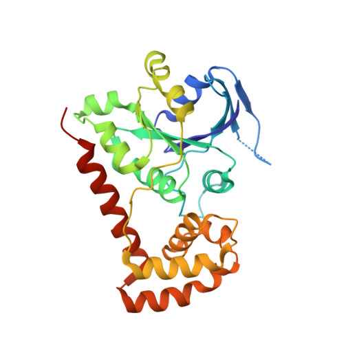
The release of GDP from GTPases signals the initiation of a GTPase cycle, where the association of GTP triggers conformational changes promoting binding of downstream effector molecules. Studies have implicated the nucleotide-binding G5 loop to be involved in the GDP release mechanism. For example, biophysical studies on both the eukaryotic Gα proteins and the GTPase domain (NFeoB) of prokaryotic FeoB proteins have revealed conformational changes in the G5 loop that accompany nucleotide binding and release. However, it is unclear whether this conformational change in the G5 loop is a prerequisite for GDP release, or, alternatively, the movement is a consequence of release. To gain additional insight into the sequence of events leading to GDP release, we have created a chimeric protein comprised of Escherichia coli NFeoB and the G5 loop from the human Giα1 protein. The protein chimera retains GTPase activity at a similar level to wild-type NFeoB, and structural analyses of the nucleotide-free and GDP-bound proteins show that the G5 loop adopts conformations analogous to that of the human nucleotide-bound Giα1 protein in both states. Interestingly, isothermal titration calorimetry and stopped-flow kinetic analyses reveal uncoupled nucleotide affinity and release rates, supporting a model where G5 loop movement promotes nucleotide release.
Organizational Affiliation:
Structural Biology Program, Centenary Institute, Sydney, New South Wales, Australia; Faculty of Medicine, Central Clinical School, University of Sydney, Sydney, New South Wales, Australia.