


Crystal structure of glucosamine-6-phosphate deaminase from Vibrio cholerae
Maltseva, N., Kim, Y., Kwon, K., Anderson, W.F., Joachimiak, A., CSGIDTo be published.
Experimental Data Snapshot
wwPDB Validation 3D Report Full Report

Entity ID: 1 | |||||
|---|---|---|---|---|---|
| Molecule | Chains | Sequence Length | Organism | Details | Image |
| Glucosamine-6-phosphate deaminase | 274 | Vibrio cholerae O1 biovar El Tor str. N16961 | Mutation(s): 0 Gene Names: nagB, VC_A1025 EC: 3.5.99.6 | 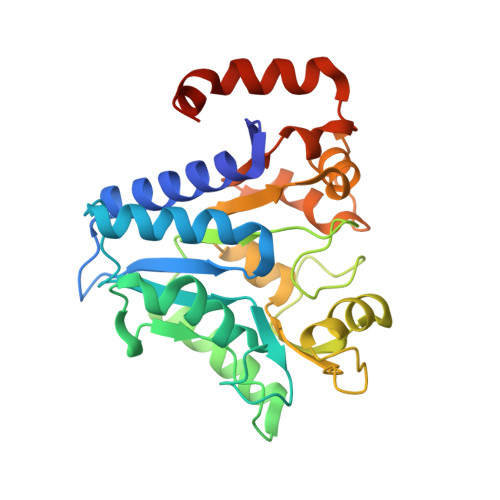 | |
UniProt | |||||
Find proteins for Q9KKS5 (Vibrio cholerae serotype O1 (strain ATCC 39315 / El Tor Inaba N16961)) Explore Q9KKS5 Go to UniProtKB: Q9KKS5 | |||||
Entity Groups | |||||
| Sequence Clusters | 30% Identity50% Identity70% Identity90% Identity95% Identity100% Identity | ||||
| UniProt Group | Q9KKS5 | ||||
Sequence AnnotationsExpand | |||||
| |||||
| Ligands 4 Unique | |||||
|---|---|---|---|---|---|
| ID | Chains | Name / Formula / InChI Key | 2D Diagram | 3D Interactions | |
| GOL Query on GOL | F [auth A] | GLYCEROL C3 H8 O3 PEDCQBHIVMGVHV-UHFFFAOYSA-N |  | ||
| FMT Query on FMT | H [auth B] | FORMIC ACID C H2 O2 BDAGIHXWWSANSR-UHFFFAOYSA-N |  | ||
| CL Query on CL | E [auth A] | CHLORIDE ION Cl VEXZGXHMUGYJMC-UHFFFAOYSA-M |  | ||
| MG Query on MG | D [auth A], G [auth B], I [auth C] | MAGNESIUM ION Mg JLVVSXFLKOJNIY-UHFFFAOYSA-N |  | ||
| Length ( Å ) | Angle ( ˚ ) |
|---|---|
| a = 78.359 | α = 90 |
| b = 135.124 | β = 106.15 |
| c = 86.001 | γ = 90 |
| Software Name | Purpose |
|---|---|
| SBC-Collect | data collection |
| HKL-3000 | data collection |
| HKL-3000 | phasing |
| MOLREP | phasing |
| PHENIX | refinement |
| HKL-3000 | data reduction |
| HKL-3000 | data scaling |
RCSB PDB (citation) is hosted by
RCSB PDB is a member of the

