


Crystal Structure of Gaf Domain of Potassium Sensor Histidine Kinase Kdpd from Escherichia Coli
Kumar, S., Yernool, D.A.To be published.
Experimental Data Snapshot
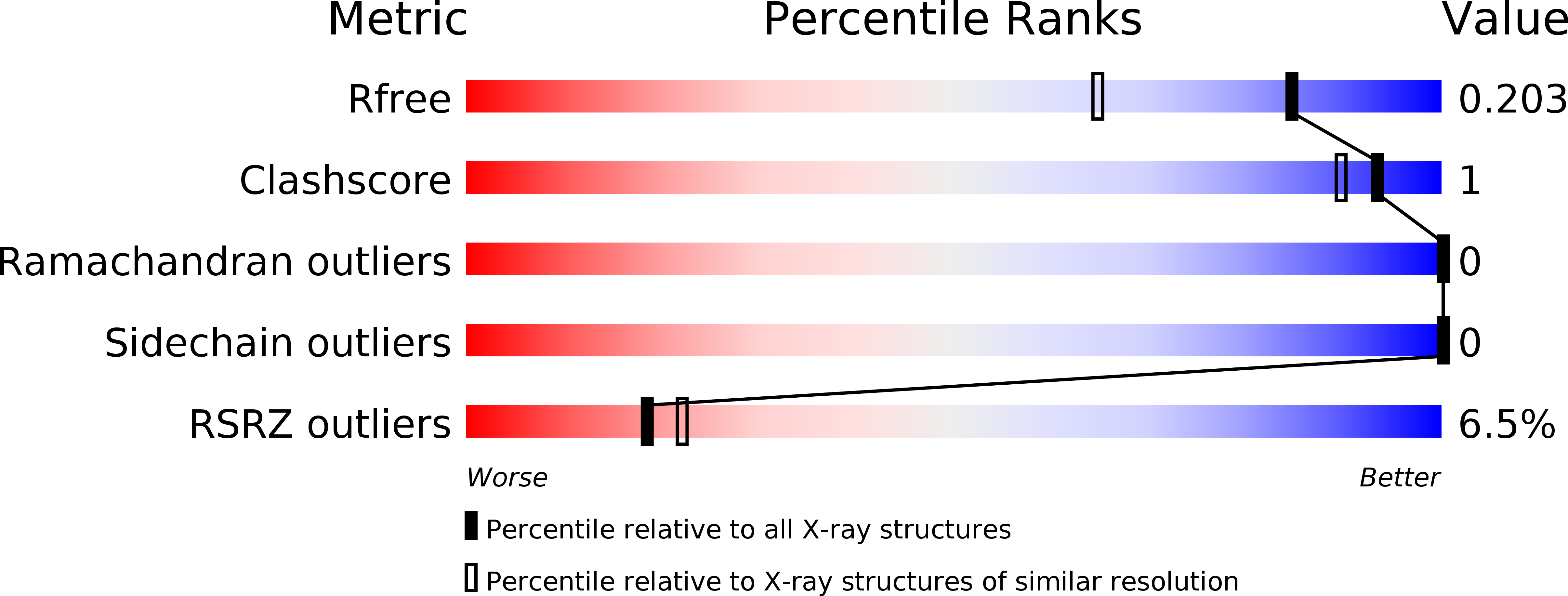
wwPDB Validation 3D Report Full Report
Entity ID: 1 | |||||
|---|---|---|---|---|---|
| Molecule | Chains | Sequence Length | Organism | Details | Image |
| Sensor protein KdpD | 149 | Escherichia coli K-12 | Mutation(s): 0 Gene Names: b0695, ECDH1ME8569_0654, EcDH1_2941, JW0683, kdpD EC: 2.7.13.3 | 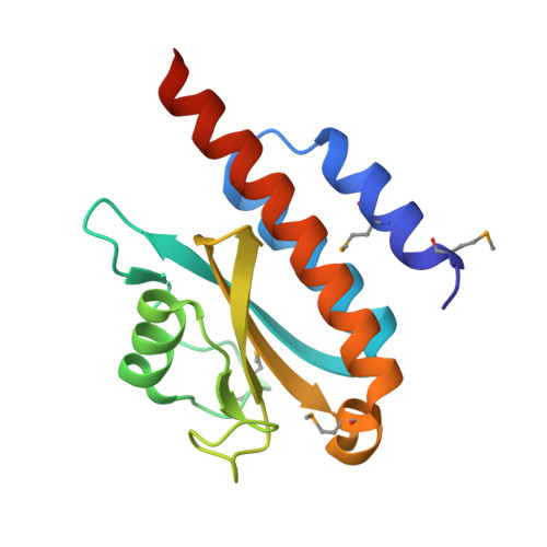 | |
UniProt | |||||
Find proteins for P21865 (Escherichia coli (strain K12)) Explore P21865 Go to UniProtKB: P21865 | |||||
Entity Groups | |||||
| Sequence Clusters | 30% Identity50% Identity70% Identity90% Identity95% Identity100% Identity | ||||
| UniProt Group | P21865 | ||||
Sequence AnnotationsExpand | |||||
| |||||
| Modified Residues 1 Unique | |||||
|---|---|---|---|---|---|
| ID | Chains | Type | Formula | 2D Diagram | Parent |
| MSE Query on MSE | A | L-PEPTIDE LINKING | C5 H11 N O2 Se |  | MET |
| Length ( Å ) | Angle ( ˚ ) |
|---|---|
| a = 35.31 | α = 90 |
| b = 57.61 | β = 90 |
| c = 61 | γ = 90 |
| Software Name | Purpose |
|---|---|
| PHENIX | model building |
| PHENIX | refinement |
| MOSFLM | data reduction |
| SCALA | data scaling |
| PHENIX | phasing |
RCSB PDB (citation) is hosted by
RCSB PDB is a member of the

