


Crystal structure of FMN quinone reductase 2 at 1.55A
Serriere, J., Boutin, J.A., Isabet, T., Antoine, M., Ferry, G.To be published.
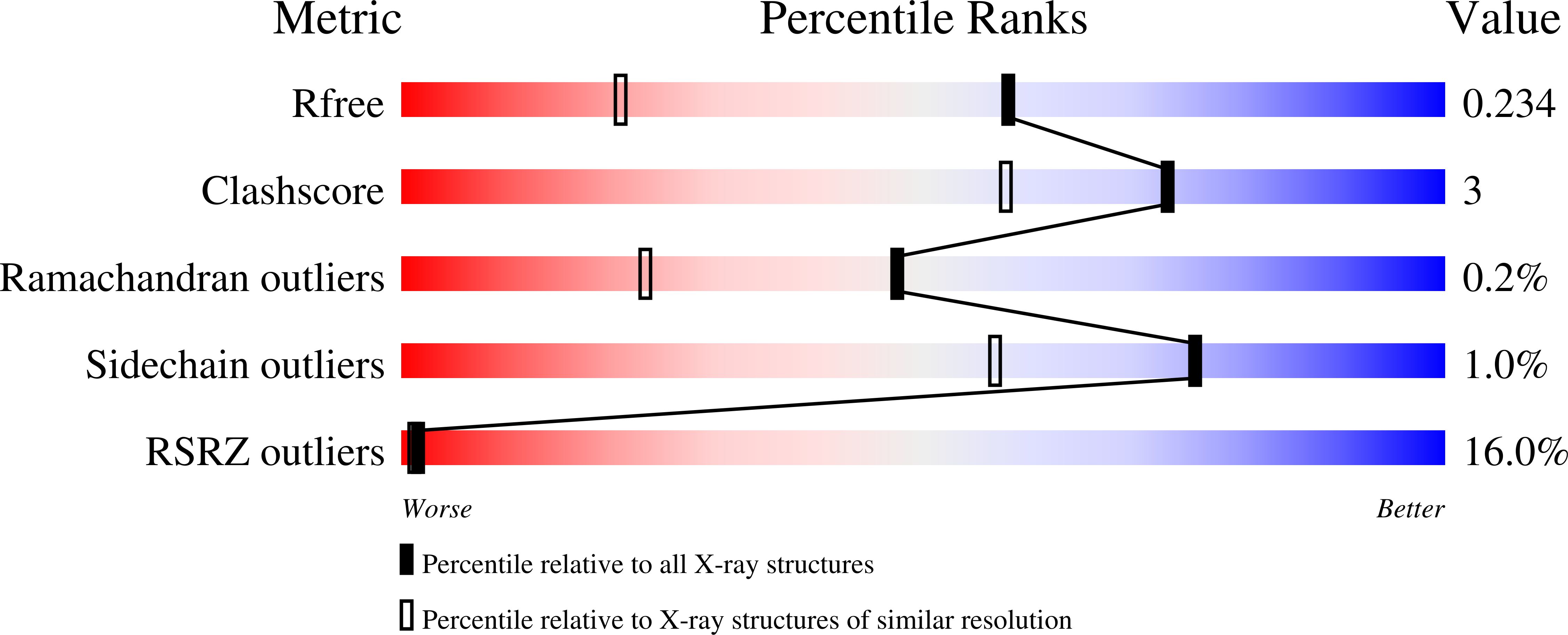
Experimental Data Snapshot
Entity ID: 1 | |||||
|---|---|---|---|---|---|
| Molecule | Chains | Sequence Length | Organism | Details | Image |
| Ribosyldihydronicotinamide dehydrogenase [quinone] | 231 | Homo sapiens | Mutation(s): 1 Gene Names: NQO2, NMOR2 EC: 1.10.99.2 | 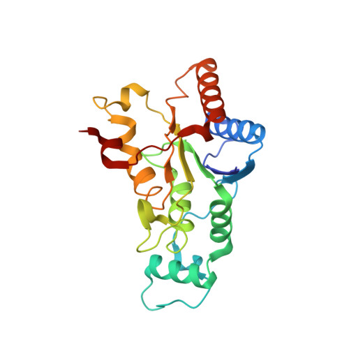 | |
UniProt & NIH Common Fund Data Resources | |||||
Find proteins for P16083 (Homo sapiens) Explore P16083 Go to UniProtKB: P16083 | |||||
PHAROS: P16083 GTEx: ENSG00000124588 | |||||
Entity Groups | |||||
| Sequence Clusters | 30% Identity50% Identity70% Identity90% Identity95% Identity100% Identity | ||||
| UniProt Group | P16083 | ||||
Sequence AnnotationsExpand | |||||
| |||||
| Ligands 2 Unique | |||||
|---|---|---|---|---|---|
| ID | Chains | Name / Formula / InChI Key | 2D Diagram | 3D Interactions | |
| FMN Query on FMN | C [auth A], E [auth B] | FLAVIN MONONUCLEOTIDE C17 H21 N4 O9 P FVTCRASFADXXNN-SCRDCRAPSA-N |  | ||
| ZN Query on ZN | D [auth A], F [auth B] | ZINC ION Zn PTFCDOFLOPIGGS-UHFFFAOYSA-N |  | ||
| Length ( Å ) | Angle ( ˚ ) |
|---|---|
| a = 56.17 | α = 90 |
| b = 83.27 | β = 90 |
| c = 106.44 | γ = 90 |
| Software Name | Purpose |
|---|---|
| XDS | data scaling |
| MOLREP | phasing |
| BUSTER | refinement |
| XDS | data reduction |
RCSB PDB (citation) is hosted by
RCSB PDB is a member of the

