


Crystal Structure of PduA with edge mutation K26D
Pang, A.H., Sawaya, M.R., Bobik, T.A., Yeates, T.O.To be published.
Experimental Data Snapshot
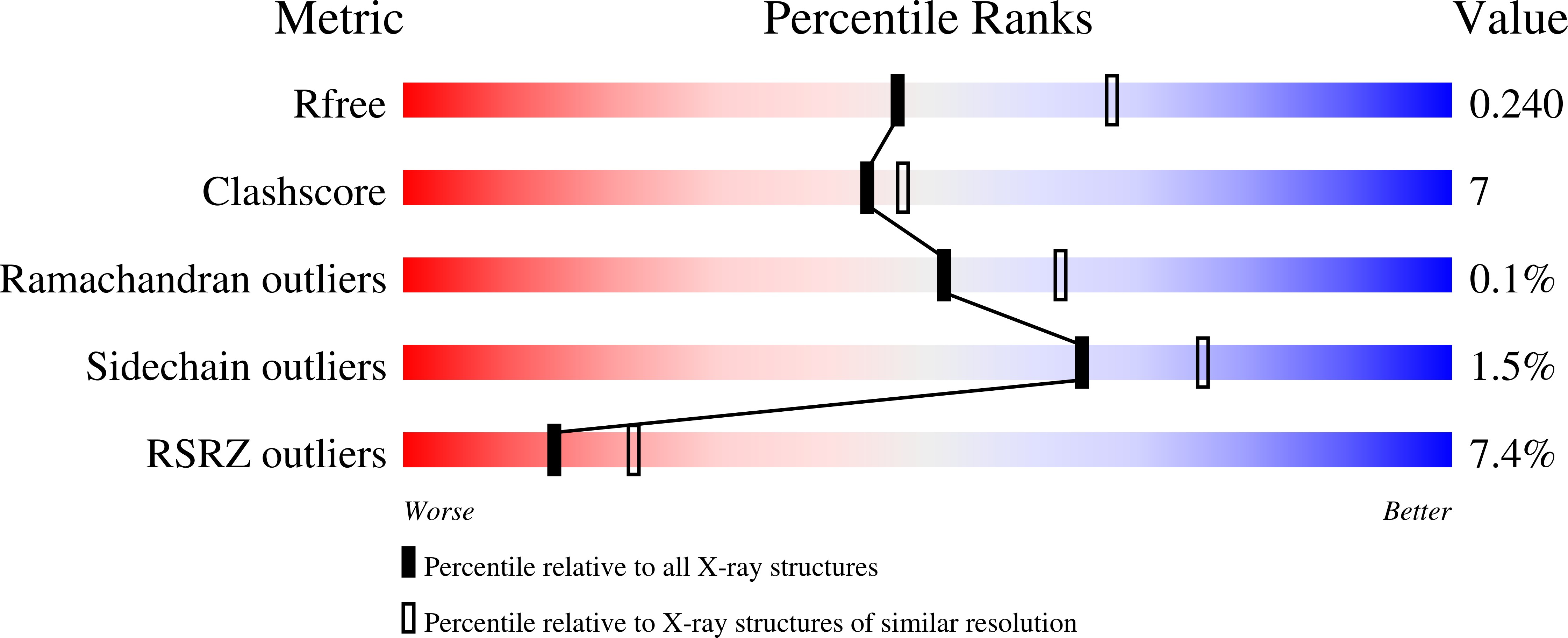
wwPDB Validation 3D Report Full Report
Entity ID: 1 | |||||
|---|---|---|---|---|---|
| Molecule | Chains | Sequence Length | Organism | Details | Image |
| Propanediol utilization protein PduA | 102 | Salmonella enterica subsp. enterica serovar Typhimurium str. LT2 | Mutation(s): 1 Gene Names: pduA, STM2038 | 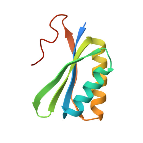 | |
UniProt | |||||
Find proteins for P0A1C7 (Salmonella typhimurium (strain LT2 / SGSC1412 / ATCC 700720)) Explore P0A1C7 Go to UniProtKB: P0A1C7 | |||||
Entity Groups | |||||
| Sequence Clusters | 30% Identity50% Identity70% Identity90% Identity95% Identity100% Identity | ||||
| UniProt Group | P0A1C7 | ||||
Sequence AnnotationsExpand | |||||
| |||||
| Ligands 2 Unique | |||||
|---|---|---|---|---|---|
| ID | Chains | Name / Formula / InChI Key | 2D Diagram | 3D Interactions | |
| SO4 Query on SO4 | K [auth A], L [auth D], N [auth H] | SULFATE ION O4 S QAOWNCQODCNURD-UHFFFAOYSA-L |  | ||
| GOL Query on GOL | J [auth A], M [auth G] | GLYCEROL C3 H8 O3 PEDCQBHIVMGVHV-UHFFFAOYSA-N |  | ||
| Length ( Å ) | Angle ( ˚ ) |
|---|---|
| a = 108.11 | α = 90 |
| b = 108.11 | β = 90 |
| c = 334.34 | γ = 120 |
| Software Name | Purpose |
|---|---|
| ADSC | data collection |
| PHENIX | model building |
| PHENIX | refinement |
| DENZO | data reduction |
| SCALEPACK | data scaling |
| PHENIX | phasing |
RCSB PDB (citation) is hosted by
RCSB PDB is a member of the

